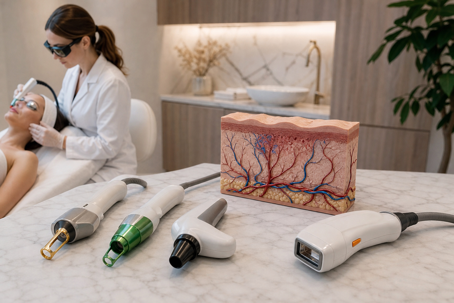
Vascular lasers represent a sophisticated advancement in dermatological and aesthetic medicine, designed to address a variety of conditions involving visible capillaries, persistent redness, and superficial vascular lesions. These treatments are especially significant for individuals experiencing facial redness, rosacea, broken capillaries, or vascular birthmarks—conditions that often detract from skin clarity and uniformity. The emergence and refinement of vascular lasers have provided practitioners with versatile tools to selectively target problematic blood vessels, thereby restoring a clearer and more balanced skin tone.
Understanding vascular lasers for capillaries and redness is essential not only for clinicians but also for patients exploring effective options for management of these common skin concerns. This article serves as a comprehensive exploration of the types of vascular lasers available, elaborating on their mechanisms of action, and offering clarity on how each device suits particular vascular issues. Such knowledge is invaluable when navigating treatment choices, as it dispels the notion of a one-size-fits-all approach and highlights the individualized strategies necessary for optimal outcomes in vascular laser therapy.
The importance of these treatments transcends aesthetic improvement; they can also alleviate psychosocial distress and improve quality of life for people affected by visible vascular lesions. Our focus here is detailed yet accessible, examining foundational concepts like selective photothermolysis alongside practical considerations in device selection, treatment planning, and patient expectations. Ultimately, the article aims to convey the nuanced sophistication of vascular lasers and how they integrate into the broader spectrum of laser procedures for skin health and appearance.
What Is Selective Photothermolysis and Why Is It Crucial in Vascular Laser Treatment?
Selective photothermolysis is the fundamental principle that underpins the effectiveness of vascular lasers. Coined by Dr. Rox Anderson and Dr. John Parrish in the 1980s, selective photothermolysis describes the process by which specific wavelengths of light are absorbed by targeted chromophores—in this case, hemoglobin within blood vessels—resulting in localized thermal injury that spares surrounding tissues.
This mechanism is pivotal in treating capillaries and redness because it allows lasers to precisely destroy abnormal or unwanted vessels without damaging the epidermis or adjacent structures. The selectivity is achieved by carefully choosing the laser wavelength, pulse duration, and energy settings that correspond to the absorption spectrum and thermal relaxation time of the targeted vessels.
When vascular lesions are exposed to laser light absorbed selectively by hemoglobin, the rapid heating causes coagulation and collapse of the blood vessel walls. The body subsequently reabsorbs these damaged vessels, leading to a gradual clearing of redness and visible capillaries. The finesse of this process minimizes downtime, reduces risk of scarring, and enhances treatment tolerability.
How Does Vascular Laser Work on Capillaries?
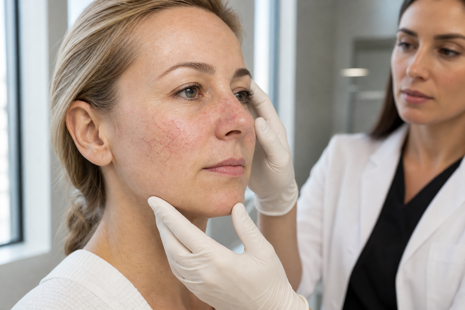
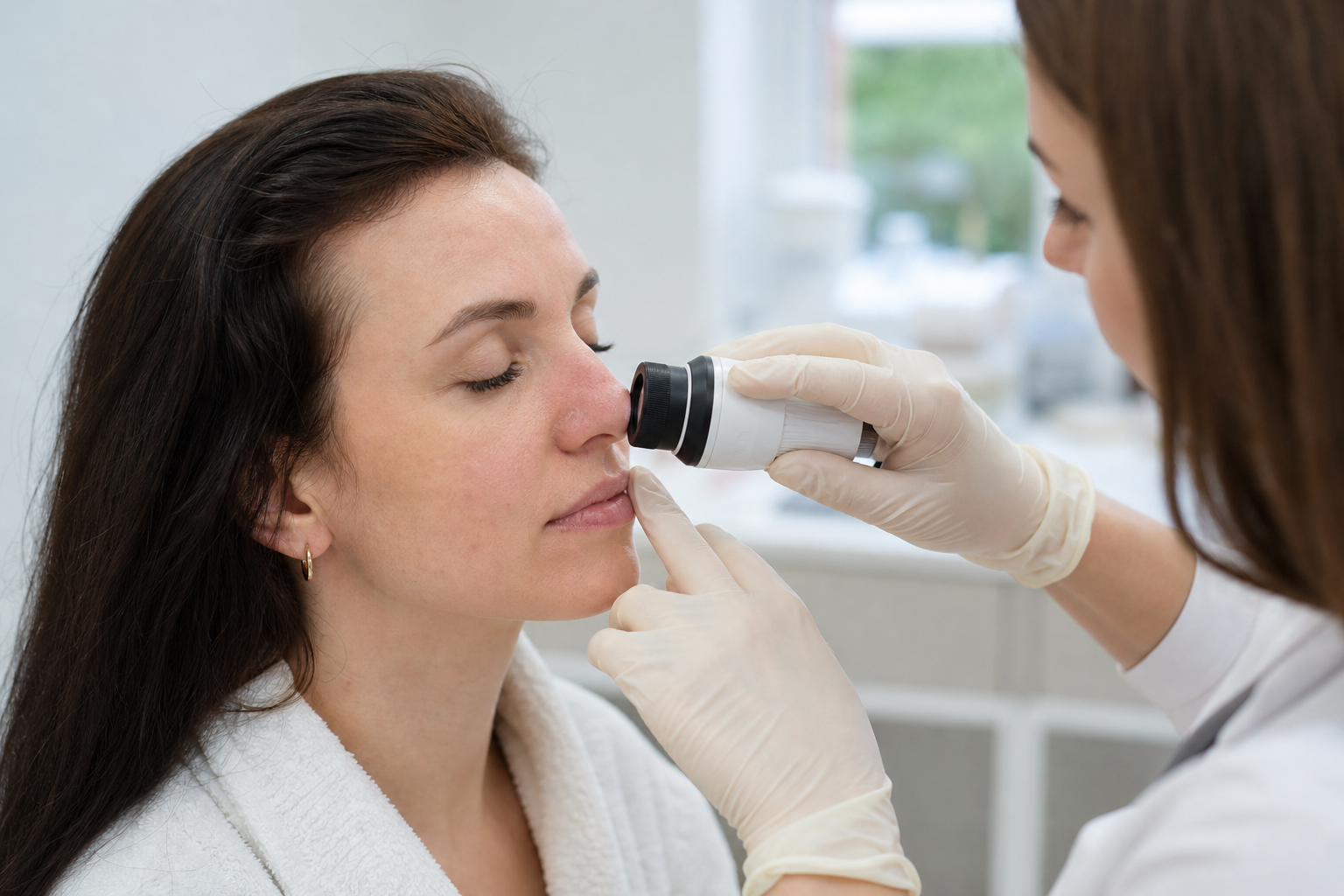
Capillaries, the smallest blood vessels in the body, can become dilated or broken due to a variety of factors including sun damage, aging, rosacea, or trauma. These changes give rise to visible blood vessels near the skin surface, creating a mottled or flushed appearance. Vascular lasers target these vessels by exploiting their absorption properties as described through selective photothermolysis.
The treatment involves directing pulsed light of a precise wavelength onto the affected areas. This wavelength is preferentially absorbed by oxyhemoglobin and deoxyhemoglobin in red blood cells, generating heat that selectively injures the vascular endothelium. The vessel walls are damaged enough to cause their collapse, while surrounding skin remains largely unharmed.
Repeated sessions are often necessary to incrementally reduce vascular visibility, as complete clearance depends on vessel size, depth, and the patient’s skin type. The ability to adjust parameters such as fluence (energy density), pulse duration, and spot size allows practitioners to customize treatments for individual capillaries or complex patterns of redness.
Pulsed Dye Laser: What Red Skin Conditions Does It Treat?
The Pulsed Dye Laser (PDL) stands as one of the most extensively studied and utilized devices in the vascular laser category. Emitting light typically around 585 to 595 nanometers, PDL is ideally absorbed by oxyhemoglobin, making it particularly effective for addressing superficial vascular lesions.
PDL is commonly employed to treat conditions such as rosacea, facial telangiectasias, port-wine stains, and poikiloderma. Its efficacy extends to reducing redness in inflammatory dermatoses and promoting improvement in hypertrophic scars and keloids through modulation of vascularity.
The unique advantages of PDL include its established safety profile and precision in targeting small to medium-sized vessels in the upper dermis. Moreover, it often results in a predictable purpura (bruising) that serves as an indicator of adequate vascular injury, guiding expectations for transient side effects and recovery.
Nd:YAG Laser for Capillaries: When Is It Used?
The Neodymium-doped Yttrium Aluminum Garnet (Nd:YAG) laser operates at a wavelength of 1064 nanometers, giving it the capability to penetrate deeper into the skin compared to other vascular lasers. This feature makes Nd:YAG particularly useful for treating larger and deeper blood vessels that are not accessible with shorter-wavelength devices.
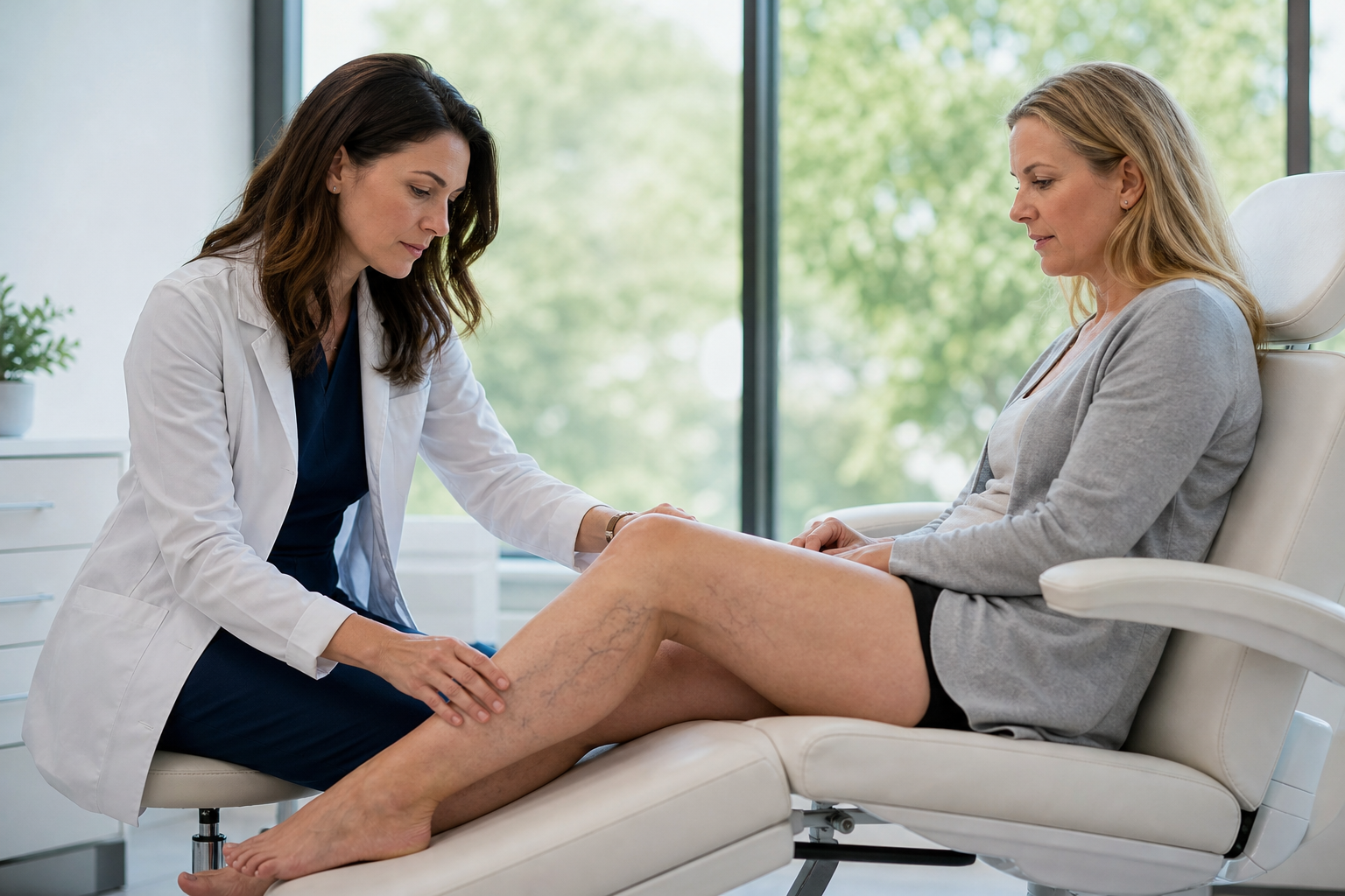
Its application includes the treatment of leg veins, reticular veins, and certain types of rosacea-associated telangiectasias that involve deeper vasculature. Despite its deeper reach, Nd:YAG laser can also be adapted to address facial capillaries, although care must be taken to avoid damaging the epidermis, particularly in patients with darker skin phototypes.
Due to its penetrating capacity, Nd:YAG is a preferred choice when other lasers fail to effectively resolve deeper vessels or when combined vascular lesions of varied depth exist. It requires meticulous parameter selection to balance efficacy and safety, emphasizing the significance of practitioner expertise.
KTP Laser for Redness and Superficial Capillaries
The Potassium Titanyl Phosphate (KTP) laser operates at a wavelength of approximately 532 nanometers, favoring the treatment of superficial cutaneous vascular lesions by virtue of its absorption characteristics. It excels in targeting red-colored vessels close to the skin’s surface, making it ideal for fine capillaries and diffuse redness.
Patients with facial redness due to conditions such as telangiectasias, rosacea-related erythema, and spider veins often benefit from KTP laser therapy. The system offers precise control and fewer side effects when administered appropriately.
KTP lasers generally cause minimal purpura, which is advantageous for individuals seeking less downtime. Their utility is somewhat limited by the penetration depth, as they are less effective on deeper or larger vessels, highlighting the importance of understanding lesion characteristics in device selection.
How IPL Works for Capillaries and Redness
Intense Pulsed Light (IPL) systems, while not lasers, employ broad-spectrum light filtered to specific wavelength ranges targeting hemoglobin and melanin. IPL delivers multiple wavelengths simultaneously, producing a photothermal effect that damages vascular structures and stimulates collagen remodeling.
IPL is valued for its versatility and ability to treat diffuse redness, erythema, pigmentation, and fine capillaries. It is frequently used for patients with rosacea and other vascular-related skin conditions who prefer non-laser options with broader treatment capabilities.
The pulsed nature and energy parameters of IPL can be adjusted to balance efficacy and safety, catering to various skin types and lesion depths. Its capacity to cover larger treatment areas quickly is advantageous in managing extensive redness but may require multiple sessions for optimal results.
IPL Is Not a Laser: What’s the Real Difference?
While both IPL and lasers are light-based therapies, their fundamental difference lies in the nature of the light emitted. Lasers produce monochromatic, coherent, and collimated light—meaning a single wavelength, tightly focused and in phase—allowing concentrated and targeted energy delivery.
In contrast, IPL uses incoherent, polychromatic, and non-collimated light. This means it emits a broad spectrum of wavelengths that can be selectively filtered to target certain chromophores but results in more dispersed energy delivery compared to lasers. This broader approach allows IPL to treat multiple concerns simultaneously but may lack the precision that lasers provide.
This distinction affects treatment outcomes, side effect profiles, and patient suitability. Some patients may benefit from IPL’s versatility and shorter treatment times, while others require the specificity and deeper penetration offered by vascular lasers.
Factors Influencing the Choice of Vascular Laser Device
Selecting the appropriate laser or light device for vascular lesions depends on a constellation of factors, including vessel color, diameter, depth, location, and patient skin phototype. No single device or set of parameters is universally superior; instead, the choice must be tailored to the lesion characteristics and patient needs.
For instance, superficial red capillaries respond well to PDL or KTP lasers due to their specific wavelengths, whereas deeper blue or purple veins may require Nd:YAG laser treatment. Skin phototype influences risk of hyperpigmentation and must guide energy settings and device selection, particularly in darker skin.
Other considerations entail patient tolerance for downtime and side effects, previous treatment history, and concurrent skin conditions that may affect healing or response. Comprehensive assessment ensures the best possible balance of efficacy, safety, and patient satisfaction.
What to Expect During and After Vascular Laser Treatment
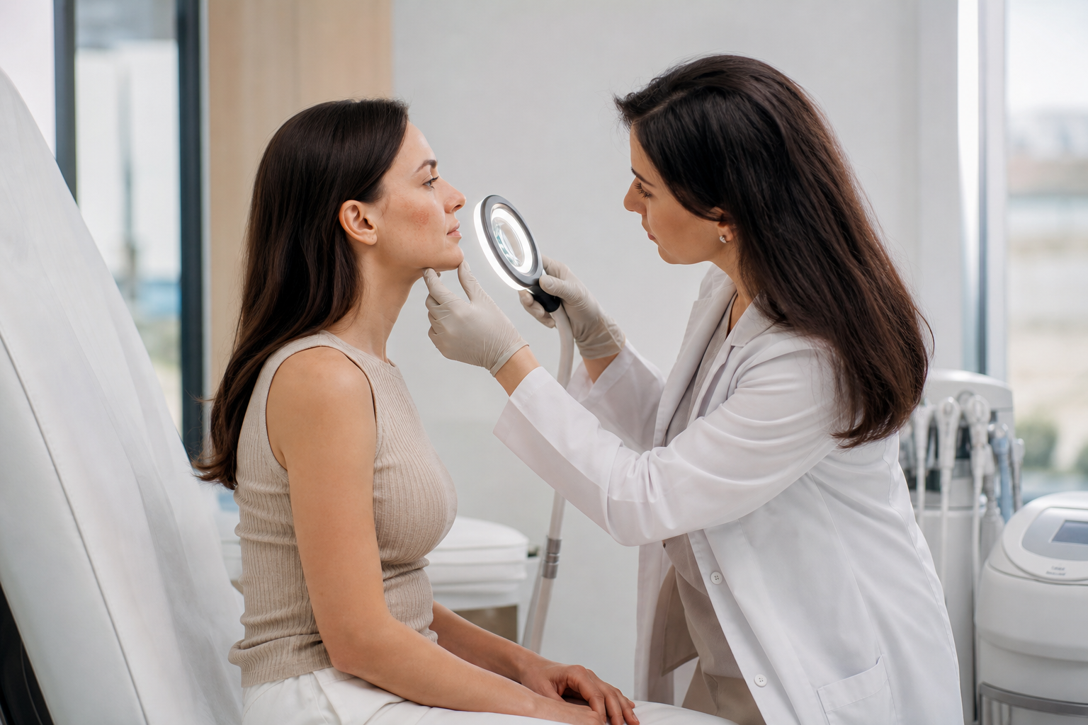
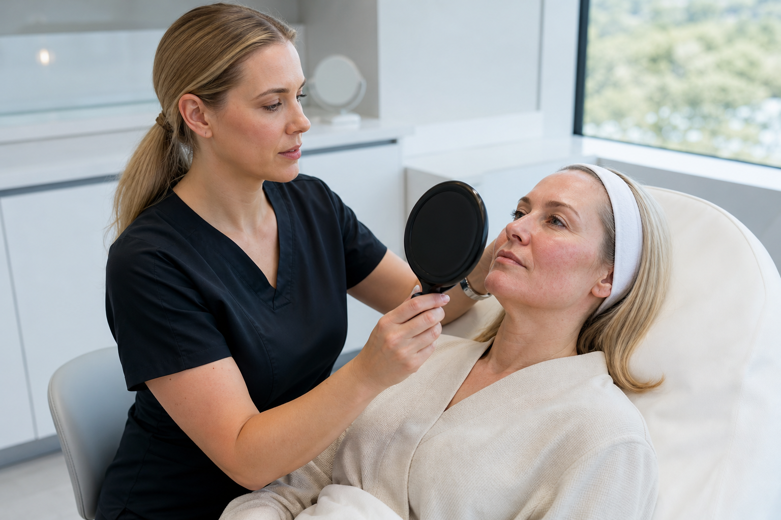
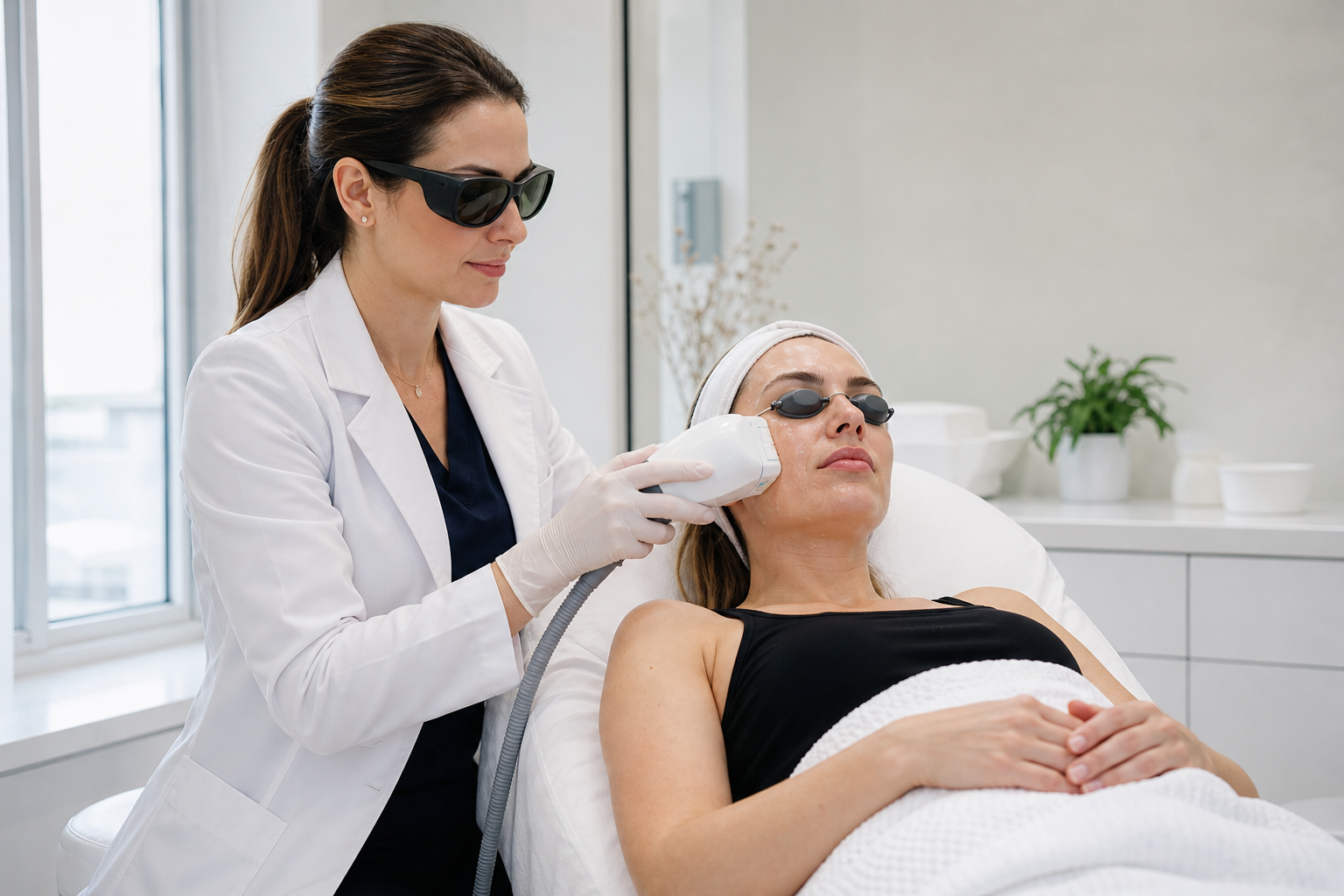
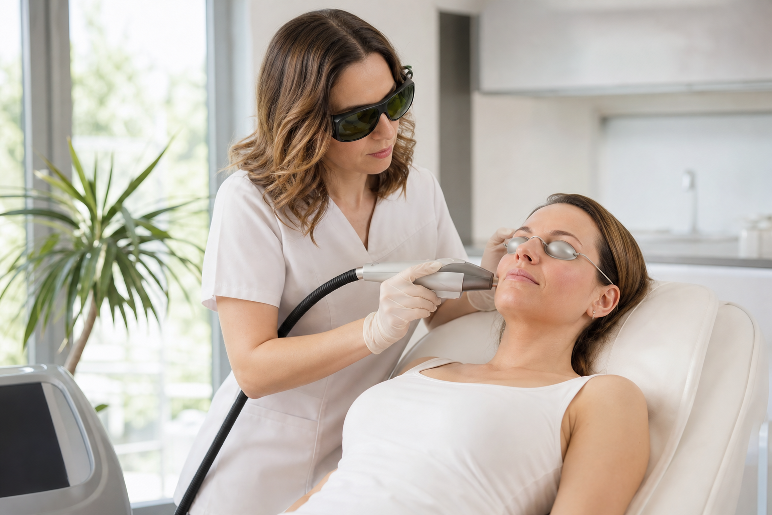
Patients undergoing vascular laser therapy can anticipate a structured treatment process beginning with an evaluation to select the optimal device and parameters. During treatment, a cooling mechanism is often used to protect the epidermis and enhance comfort.
The sensation is typically described as a brief snapping or warmth. Post-treatment effects may include mild erythema, swelling, and in some cases, purpura or crusting, especially with PDL. These effects are generally transient, resolving within days to weeks depending on the device used.
Multiple treatment sessions spaced several weeks apart are typically needed to achieve significant improvement. Maintenance treatments may be recommended to sustain results. Patient adherence to sun protection and skincare instructions enhances outcomes and reduces risk of complications.
Addressing Common Questions About Vascular Laser Treatments
Many patients inquire about pain levels, number of sessions, potential side effects, and expected longevity of results. While individual experiences vary, vascular laser treatments are generally well tolerated and safe when performed by experienced clinicians.
Side effects are usually mild and temporary but can include bruising, swelling, pigmentation changes, or very rarely scarring. The number of needed sessions depends on lesion severity and response, with most requiring between three to six treatments for noticeable clearance.
Results can be long-lasting yet are subject to potential recurrence if underlying factors persist, such as sun exposure or hormonal influences. Comprehensive care and periodic touch-ups help maintain improvements over time.