The appearance of red skin lesions can be distressing, not only because they affect the visible aesthetics of the skin but also due to the potential underlying vascular abnormalities they indicate. Pulsed dye laser (PDL) therapy has become a cornerstone in dermatological treatment options for managing various red skin lesions. This advanced laser treatment selectively targets aberrant blood vessels, offering precise, effective solutions for patients seeking clearer, more even skin tone. Understanding which red skin lesions respond best to PDL is essential for clinicians, patients, and anyone interested in dermatological laser treatments.
Unlike other light-based treatments such as intense pulsed light (IPL), which is often conflated with laser therapy, pulsed dye laser works through a specific mechanism called selective photothermolysis. This ability to target vascular structures at specific wavelengths distinguishes it from broad-spectrum devices and makes it highly effective for vascular lesions.
This article is crafted for dermatologists, clinicians, and patients exploring treatment options for vascular skin concerns. It outlines the types of red skin lesions treatable by pulsed dye laser and explains crucial factors that influence treatment choice, including lesion color, vessel diameter, depth, location, and skin phototype. In doing so, it highlights the sophisticated science underpinning PDL therapy and sets realistic expectations about outcomes.
What Is Pulsed Dye Laser and How Does It Work for Vascular Skin Lesions?
Pulsed dye laser is a type of vascular laser treatment designed to target and treat blood vessels located close to the skin surface. The device emits a concentrated beam of light at approximately 585 to 595 nanometers, a wavelength highly absorbed by oxyhemoglobin, the oxygen-carrying pigment in red blood cells.
When the light energy is absorbed by the targeted blood vessel, it converts to heat, causing selective damage to the vessel walls without significantly affecting the surrounding tissue. This process, known as selective photothermolysis, allows for the precise eradication of abnormal or dilated blood vessels that manifest as red skin lesions.
The technology behind PDL ensures a carefully timed pulse of energy, minimizing collateral damage and discomfort. The laser’s parameters can be adjusted to accommodate different lesion characteristics such as size, depth, and vascularity.
Which Types of Red Skin Lesions Are Typically Treated with Pulsed Dye Laser?
Pulsed dye laser is versatile in its capability to address a range of red skin lesions rooted in vascular abnormalities. The most common indications include:
1. Port-Wine Stains (Capillary Malformations): These congenital vascular malformations appear as pink to dark red patches, commonly on the face and neck. PDL is the treatment of choice for reducing the intensity and size of port-wine stains by targeting superficial capillaries.
2. Hemangiomas: These benign vascular tumors, often occurring in infancy, can be effectively treated with pulsed dye laser during their proliferative and involuting phases to improve appearance and reduce residual redness.
3. Telangiectasias and Spider Veins: Dilated superficial blood vessels causing visible redness or web-like patterns can be managed with PDL to diminish their prominence and restore skin clarity.
4. Rosacea-Related Redness: Patients experiencing persistent facial erythema and visible blood vessels from rosacea may find symptomatic relief and cosmetic improvement through PDL treatments targeting the dilated capillaries.
5. Venous Lakes and Venous Malformations: Although larger or deeper venous lesions may require different modalities, some superficial venous lakes respond well to pulsed dye laser therapy.
How Does PDL Compare to Other Vascular Laser Treatments Like Nd:YAG and KTP Lasers?
Understanding how pulsed dye laser differs from other vascular lasers is key to selecting the optimal treatment for specific lesions. While all these lasers target blood vessels, their wavelengths and tissue penetration depths vary, influencing their suitability.
Nd:YAG Laser: Operating at a longer wavelength of 1064 nanometers, Nd:YAG laser penetrates deeper into the dermis, making it better suited for treating larger, deeper blood vessels such as thicker varicose veins or deeper venous malformations. It is also used when skin phototype is darker as it minimizes epidermal damage.
KTP Laser: With a wavelength of 532 nanometers, KTP laser primarily targets superficial blood vessels and redness. It is effective for treating superficial capillaries on the face but lacks the flexibility of pulsed dye laser for a broad range of vascular lesions.
In contrast, pulsed dye laser’s wavelength is optimal for superficial to mid-dermal capillaries and smaller vessels, allowing for effective treatment of delicate vascular lesions with precise energy delivery and minimal side effects.
Why Is IPL Not Considered a Laser and How Does This Affect Treatment Choices for Red Skin Lesions?
Intense pulsed light (IPL) devices emit a broad spectrum of light wavelengths rather than a single monochromatic wavelength like lasers. This broad range enables IPL to target multiple chromophores, including melanin and hemoglobin, making it a versatile treatment for pigmentation, vascular lesions, and skin rejuvenation.
However, IPL is not categorized as a laser because it lacks the coherent and monochromatic nature of laser light. This distinction matters when treating red skin lesions because the selective photothermolysis principle depends on precise wavelength targeting to minimize damage to surrounding tissues while effectively destroying the vascular lesion.
IPL’s broader light spectrum may reduce its specificity for certain vascular lesions when compared to PDL, which offers targeted energy delivery to oxyhemoglobin. Therefore, lesions with well-defined vascular characteristics often respond better to pulsed dye laser.
How Do Characteristics Such as Color, Diameter, Depth, and Location Influence PDL Treatment Outcomes?
The effectiveness of pulsed dye laser treatment for red skin lesions depends significantly on detailed lesion assessment. Key factors include:
Color: The color intensity of a vascular lesion correlates with the concentration and oxygenation of blood within the vessels. Bright red lesions with high oxyhemoglobin content absorb laser energy more effectively, making treatment more successful. Lesions with deeper purple or blue hues may indicate deoxygenated blood or larger vessels, which might require alternative approaches.
Diameter of Blood Vessels: Smaller vessels ranging from 0.1 to 1.0 millimeters in diameter generally respond better to PDL because the laser pulse duration and wavelength are optimized for such vessel sizes. Larger vessels may necessitate lasers with longer wavelengths and pulse durations.
Depth of Involvement: PDL targets superficial to mid-dermal vessels effectively. Deeper vascular lesions might not absorb sufficient laser energy for meaningful treatment response, or the epidermis may absorb excessive energy, increasing risk.
Location on the Body: Differences in skin thickness and vascularity across body sites impact treatment efficacy and safety. Areas with thinner skin or delicate tissue require careful parameter adjustment to avoid adverse effects such as bruising or pigmentation changes.
How Does Skin Phototype Affect Pulsed Dye Laser Treatment Selection and Settings?
Skin phototype, classified by the Fitzpatrick scale, reflects the skin’s response to ultraviolet exposure and melanin content. Higher phototypes (darker skin tones) contain more melanin, which competes with hemoglobin for laser energy absorption, potentially increasing the risk of epidermal injury and pigmentary changes.
PDL treatment for darker skin phototypes must be approached cautiously with adjusted settings such as lower fluence, longer pulse durations, and cooling techniques to protect the epidermis. While PDL remains useful, other laser modalities or treatment plans might be required depending on lesion and patient characteristics.
What Can Patients Expect During and After Pulsed Dye Laser Treatment for Red Skin Lesions?
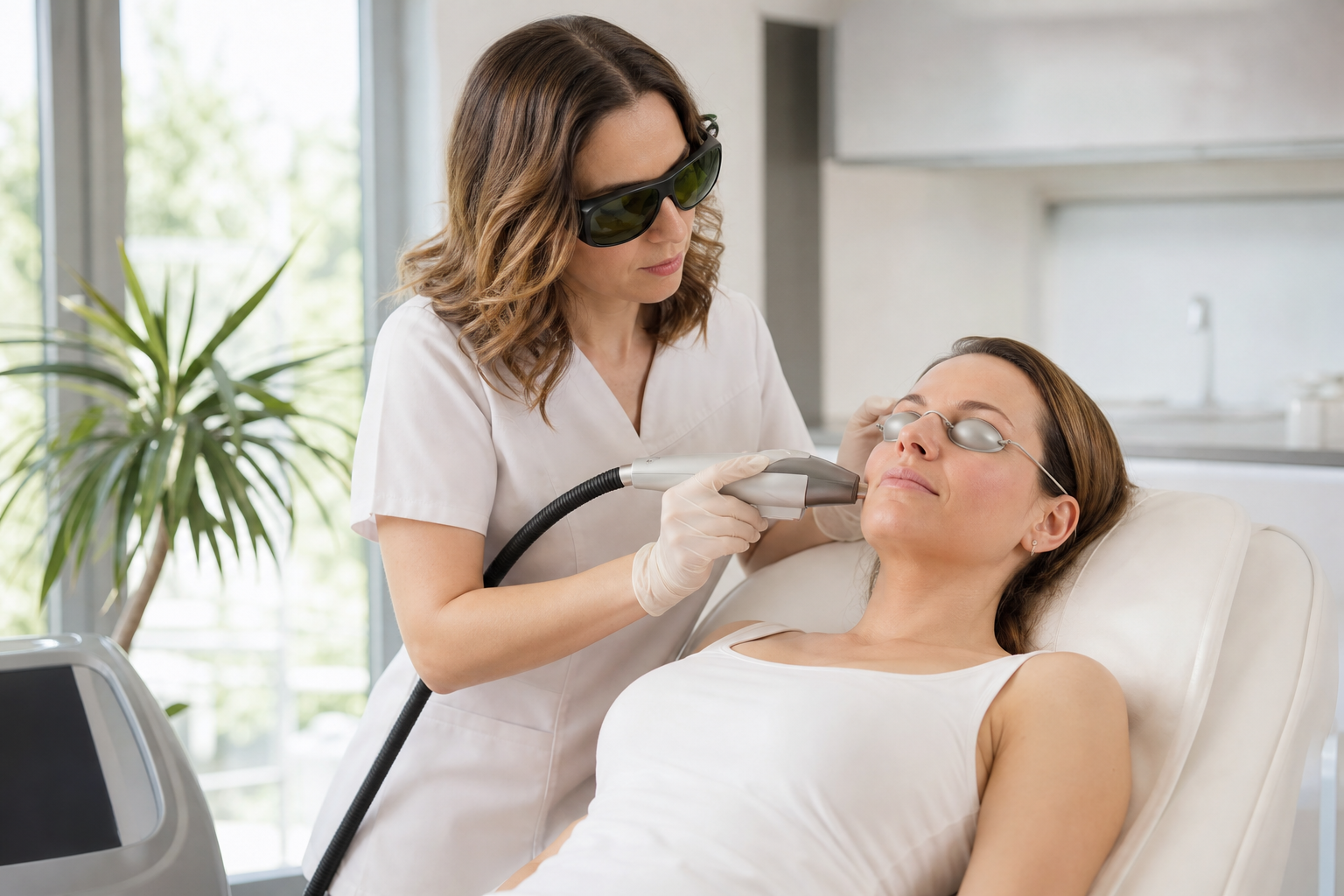
PDL treatments typically involve short pulses of laser light lasting milliseconds. During the procedure, patients may experience a sensation comparable to a rubber band snap on the skin. Topical anesthetics or cooling devices are commonly used to enhance comfort.
Immediately after treatment, the skin may appear red and swollen, sometimes with mild bruising or purpura depending on the intensity of the therapy. These side effects are usually transient and resolve within days to weeks. Following treatment, avoiding sun exposure and using gentle skin care can optimize healing.
Multiple treatment sessions are often necessary to achieve significant improvement, especially for more extensive or deeper lesions. Results include diminished redness, reduced vessel visibility, and a more uniform skin tone.
How Should Clinicians Decide Between Pulsed Dye Laser and Other Treatment Options for Red Skin Lesions?
Choosing between pulsed dye laser and other vascular laser treatments requires comprehensive evaluation of the lesion's characteristics and patient factors. Clinicians assess vessel size, depth, color, anatomical location, skin phototype, and patient preferences.
Pulsed dye laser is the preferred first-line treatment for superficial capillary malformations, small telangiectasias, and pediatric vascular birthmarks due to its specificity and safety profile. Nd:YAG or KTP lasers may offer advantages for deeper, larger vessels or darker skin phototypes.
This decision-making process benefits from evidence-based guidelines and experience with multiple laser modalities to tailor treatments achieving the best cosmetic and clinical outcomes.
What Are the Limitations and Risks Associated with Pulsed Dye Laser Therapy for Red Skin Lesions?
Despite its efficacy, pulsed dye laser has limitations. Some lesions do not respond completely and may require adjunctive treatments. Recurrence or partial clearance is possible, especially with deeper or extensive vascular malformations.
Side effects, although generally mild, can include temporary bruising, swelling, hyperpigmentation, hypopigmentation, and rarely scarring. Strict adherence to patient selection criteria and laser parameters reduces these risks.
Additionally, PDL requires multiple treatment sessions and patient commitment to follow-up schedules. It is not suitable for all vascular lesions, demanding thorough diagnosis and consultation.