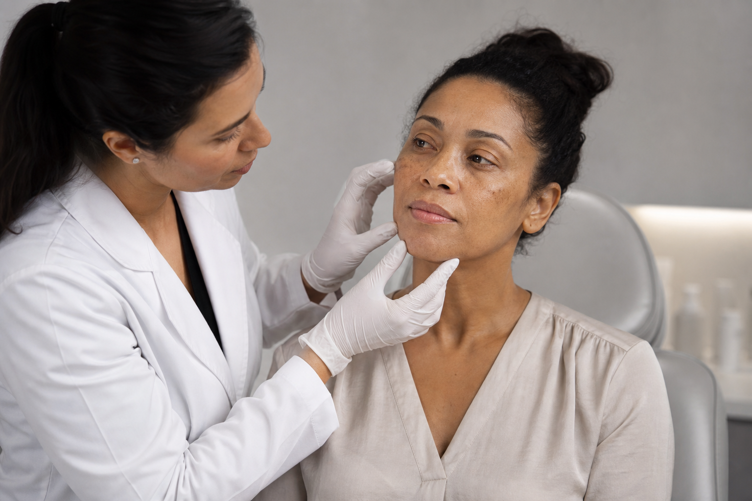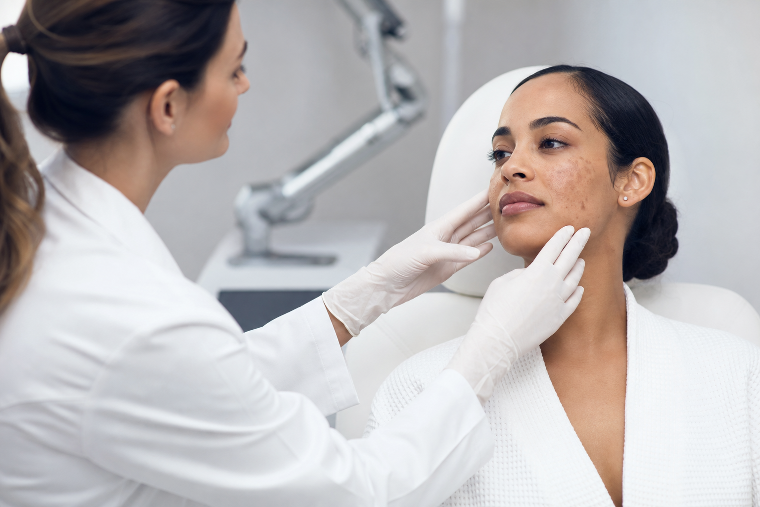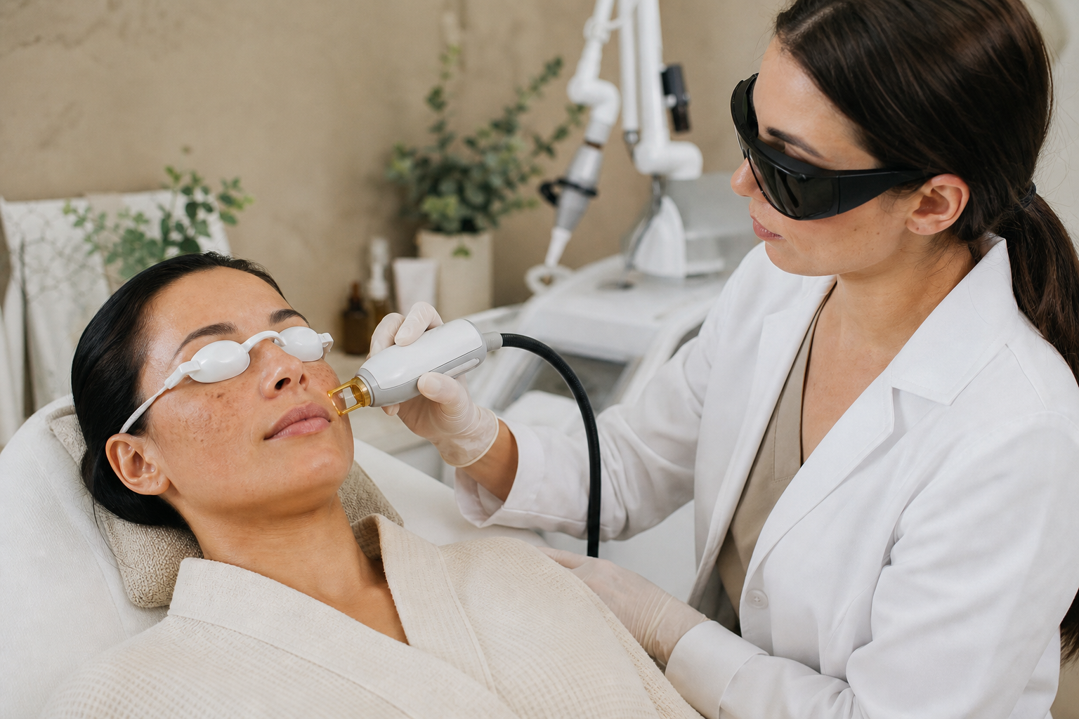
Laser pigmentation treatments have emerged as a revolutionary approach in aesthetic dermatology, offering targeted solutions for hyperpigmentation irregularities such as dark spots, melasma, and uneven skin tone. This advanced technology enables clinicians to selectively break down excess melanin deposits, promoting a more uniform complexion and enhancing skin clarity. However, while laser treatments promise effective pigmentation correction, their safety profile varies significantly based on numerous factors. Understanding these factors is crucial for dermatologists, aesthetic practitioners, and patients alike to ensure both efficacy and safety.
At the heart of laser pigmentation therapy lies a sophisticated interaction between laser technology and individual patient characteristics, most notably the Fitzpatrick skin phototype, skin condition, and pigmentation depth. The choices made regarding laser type, wavelength, and treatment parameters directly influence the risk of complications. Such complications can range from mild transient redness to more serious issues like post-inflammatory hyperpigmentation (PIH), hypopigmentation, burns, or scarring. Therefore, a well-informed treatment strategy that includes careful patient evaluation and parameter customization is essential.
This editorial aims to provide a comprehensive exploration of the safety considerations surrounding laser pigmentation treatments. We will delve into the significance of skin typing, detail the selection of appropriate laser devices and settings, examine contraindications and necessary preparations such as test spots, and discuss common risks and how to distinguish expected reactions from warning signs needing medical intervention. Through this detailed analysis, the article serves as a valuable resource for practitioners seeking to optimize patient outcomes and for individuals considering laser pigmentation therapies.
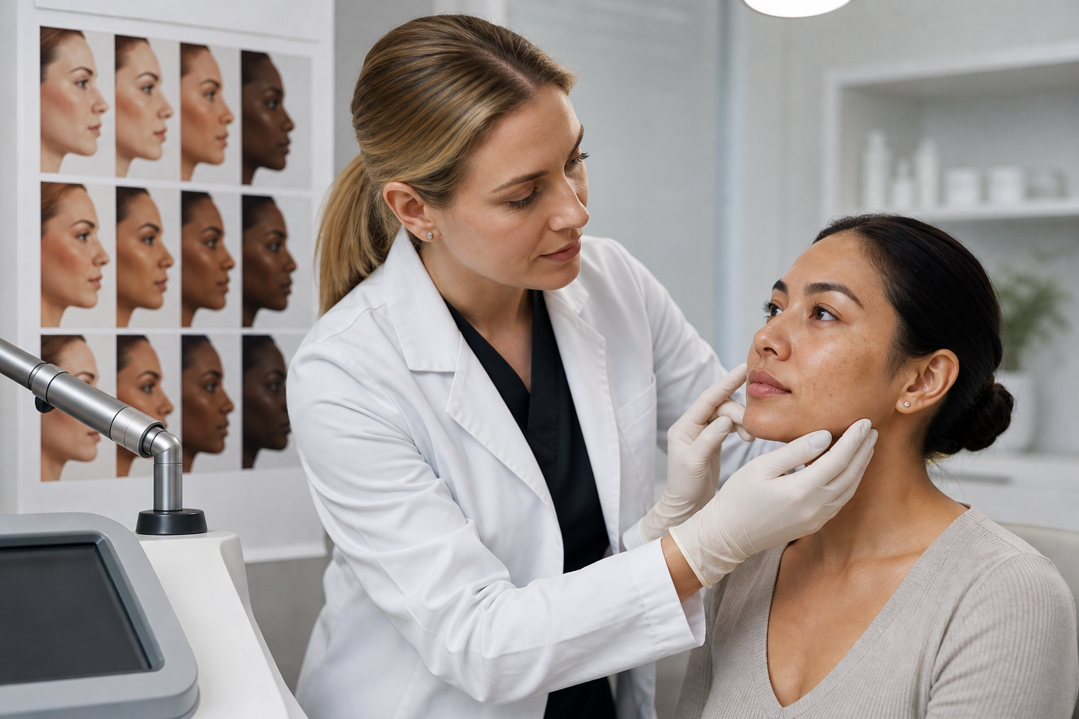
What Is Fitzpatrick Phototype and How Does It Impact Laser Pigmentation Treatment?
The Fitzpatrick phototype classification is a widely recognized system categorizing skin based on its response to ultraviolet (UV) exposure, tanning ability, and propensity for sunburn. It ranges from Type I, characterized by very fair skin that always burns and never tans, to Type VI, which denotes deeply pigmented dark skin that never burns. This classification is pivotal in tailoring laser pigmentation treatments because it directly correlates with melanin concentration and distribution within the skin.
For lighter phototypes (I–III), laser treatments typically carry a lower risk of adverse pigmentary changes, as their melanin content is comparatively low. Lasers with wavelengths that absorb efficiently in melanin, such as Alexandrite (755 nm), can be employed with relative safety and high efficacy. Conversely, higher phototypes (IV–VI) have abundant epidermal melanin, which competes with targeted pigmented lesions for laser energy absorption. This interplay increases the risk of epidermal injury manifesting as burn wounds, PIH, or hypopigmentation if inappropriate laser parameters are utilized.
In practical terms, treating darker skin types demands lasers that penetrate deeper with reduced epidermal absorption, such as the Q-Switched Nd:YAG laser at 1064 nm. This wavelength is less absorbed by epidermal melanin, minimizing thermal injury to superficial layers while effectively targeting dermal or epidermal pigmentation anomalies. Hence, assessing Fitzpatrick phototype forms the foundation for device selection and parameter adjustment, ultimately shaping the safety and success profile of pigmentation laser therapies.
Laser Technologies and Parameter Selection for Different Skin Types
Laser technology has evolved to include a variety of devices, each with unique wavelengths and pulse durations tailored to address pigmentation concerns. A foundational understanding of how these lasers interact with skin chromophores underpins safe and effective treatment delivery.
Q-Switched Nd:YAG Laser (1064 nm): Favored for darker skin types, this laser emits near-infrared light that penetrates deeply without significant epidermal absorption. Its nanosecond pulse duration allows efficient fragmentation of melanin granules with minimal collateral thermal damage, reducing PIH risk.
Alexandrite Laser (755 nm): Exhibiting high melanin absorption, this laser is effective for lighter phototypes but poses a higher burn and PIH risk in darker skin. Its shorter wavelength is absorbed primarily in superficial layers, necessitating caution in phototypes IV and above.
Diode Laser: Operating around 800-810 nm, diode lasers offer intermediate penetration and are occasionally utilized for pigmentation, though their primary applications tend toward hair removal and vascular treatments.
CO2 Laser (10,600 nm): This ablative laser vaporizes water-containing tissues and is used mainly for resurfacing rather than selective pigmentation targeting. Due to its broad tissue effect, it carries a higher risk of scarring and PIH, particularly in darker skin.
Parameter selection—including energy fluence, pulse duration, spot size, and repetition rate—is equally vital. High fluences amplify treatment efficacy but increase heat generation and the risk of burns. Long pulse durations may cause excess thermal diffusion, harming non-target tissues. Optimal spot size balances precision and treatment speed while influencing energy density. These parameters must be calibrated individually, considering skin type, pigmentation depth, and lesion characteristics.
How Do Darker and Tanned Skin Affect Laser Pigmentation Treatment Safety?
The presence of a higher melanin concentration in darker skin and tanned skin profoundly impacts laser treatment safety. Melanin acts as a broadband absorber, competing with pigmented lesions for laser energy and amplifying epidermal heating risks.
In tanned skin, melanin is temporarily increased due to UV exposure. Proceeding with laser treatment on such skin elevates the likelihood of epidermal thermal injury because the laser energy intended for pathological pigmentation is indiscriminately absorbed by enhanced background melanin. This can precipitate adverse outcomes like PIH or even blistering and scarring.
Darker skin types inherently carry this elevated risk due to melanin density. Consequently, clinicians must exercise prudence by selecting longer wavelength lasers, lowering fluence settings, and increasing pulse durations to minimize epidermal injury. Additionally, delaying treatments until tanning subsides and performing test spots becomes imperative to evaluate skin tolerance.
Ignorance of these considerations can lead to significant complications, undermining treatment efficacy and patient satisfaction. Therefore, an intimate understanding of endogenous pigmentation status and modulating treatment approach accordingly is fundamental to therapy safety.
What Are the Main Contraindications and Preparatory Steps for Lasers in Pigmentation Treatment?
Before initiating any laser procedure targeting pigmentation, practitioners must identify contraindications that could jeopardize safety or outcomes. These include but are not limited to:
Active skin infections or inflammatory dermatoses: Lasers may exacerbate inflammation or spread infection.
Herpes simplex virus outbreaks: Present a risk of viral activation post-laser trauma.
Pregnancy and breastfeeding: Safety data are limited; treatment is generally deferred.
Photosensitizing medications: Drugs like tetracyclines, retinoids (especially oral isotretinoin), and certain antidepressants increase phototoxic risks and predispose to adverse reactions.
Autoimmune diseases or coagulation disorders: Increase risks of poor healing or excessive bleeding.
Preparatory steps involve thorough patient history and medication review, physical examination, and sometimes laboratory workup. Additionally, patients should discontinue topical agents such as retinoids, hydroquinone, and alpha-hydroxy acids at least one to two weeks prior to treatment to reduce skin sensitivity.
Performing a laser test spot on a discreet skin area is also critical. This practice helps gauge individual skin response, enabling parameter adjustment and mitigating the likelihood of severe reactions. It offers a practical mechanism to anticipate pigmentation changes and inflammatory responses and tailor the main treatment accordingly.
Which Medications and Active Ingredients Affect the Safety of Laser Pigmentation Treatment?
Medication history profoundly influences laser treatment safety and should be carefully considered by clinicians. Agents that heighten cutaneous photosensitivity can drastically elevate adverse event risks.
Oral retinoids (e.g., isotretinoin): Notoriously increase epidermal fragility and impair healing. Their use requires cessation for 6 to 12 months before laser procedures.
Photosensitizing antibiotics: Such as tetracyclines and fluoroquinolones, amplify sensitivity to laser-induced injury.
Topical agents: Ingredients like tretinoin, hydroquinone, and certain acids increase epidermal turnover and may thin the skin, predisposing to irritation and burns post-laser.
Anticoagulants and antiplatelet drugs: Elevate bleeding risks and complicate healing;
Prior to initiating treatment, practitioners should coordinate with patients regarding medication discontinuation schedules, ensuring a safe therapeutic window. Constant communication aids in minimizing complications associated with these active substances.
What Are the Most Common Risks and Complications Associated with Laser Pigmentation Treatments?
Despite meticulous planning, laser pigmentation treatments carry inherent risks, the most prevalent being pigmentary alterations and physical skin damage.
Post-Inflammatory Hyperpigmentation (PIH): Following cutaneous inflammation, melanocyte stimulation leads to localized melanin overproduction, manifesting as darker patches at the treatment site. PIH is particularly common in darker skin types and can be persistent if not addressed promptly.
Hypopigmentation: Occurs due to melanocyte destruction from excessive laser energy or thermal injury, resulting in light spots or depigmented patches. This side effect is often more challenging to reverse and represents a significant aesthetic concern.
Burns and Scarring: These physical injuries arise from inappropriately high laser fluences or misapplication on sensitive or inflamed skin, potentially leading to permanent textural changes and fibrosis.
Other possible complications include erythema, edema, blistering, infection, and prolonged erythema requiring medical management. The incidence of such unwanted effects underscores the importance of stringent safety protocols and practitioner expertise.
How to Differentiate Expected Skin Reactions from Signs Requiring Medical Attention?
Post-treatment skin responses range from mild and self-limiting to severe and requiring intervention. Normal reactions typically include transient erythema, mild swelling, warmth, and a tingling sensation at the treatment site. These usually resolve within days without sequelae.
Temporary darkening of pigmented spots or light crusting before resolution is also anticipated. Conversely, certain manifestations warrant medical evaluation:
Persistent or worsening redness and swelling beyond one week;
Formation of ulcers, open wounds, or crusted lesions;
Symptoms of infection, such as pustules, increased pain, oozing, or systemic fever;
Unusual pigmentary changes, including rapidly enlarging hypopigmented or hyperpigmented areas;
Signs of scarring or textural skin distortion.
Timely recognition and intervention are critical to limit complications and facilitate recovery. Patients should receive clear post-treatment instructions detailing normal versus abnormal reactions to empower prompt reporting.