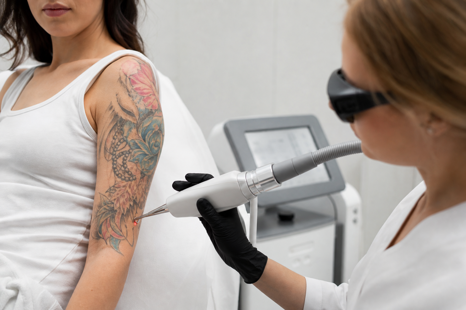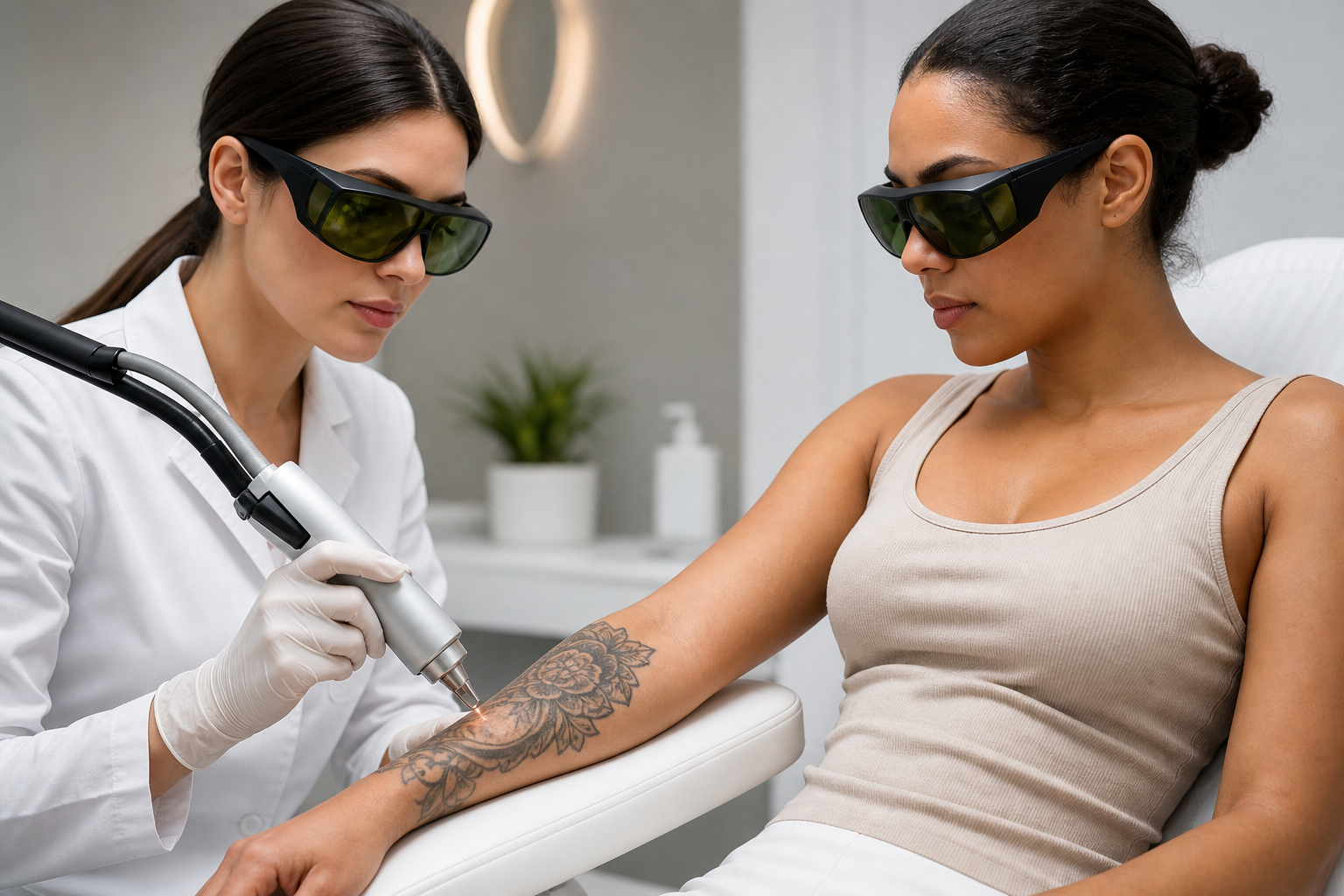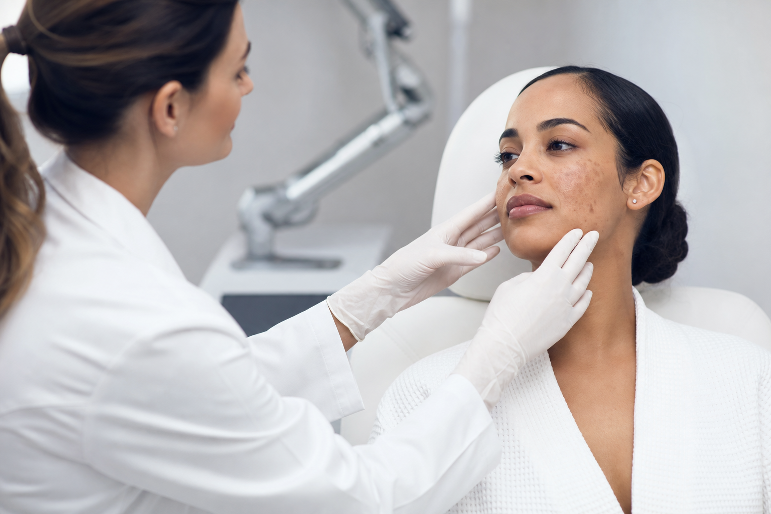
Post-inflammatory hyperpigmentation (PIH) following laser treatment represents a significant concern for both patients and dermatology professionals engaged in aesthetic and medical laser interventions. This condition involves the development of darkened skin patches or localized hyperpigmentation in areas subjected to laser therapy, potentially compromising the cosmetic outcomes and patient satisfaction achieved through these advanced procedures.
This article is crafted for individuals considering laser treatments aiming to correct pigmentation irregularities, as well as for clinicians seeking to deepen their understanding of safety aspects in laser interventions. Given the complexity imposed by the variety of laser devices, individual skin types categorized by the Fitzpatrick scale, and nuanced patient-specific factors, a comprehensive exploration of PIH is crucial. Such insight highlights the importance of tailored treatment plans, vigilant risk assessment, and prevention strategies, particularly for patients with darker or recently tanned skin.
Understanding post-inflammatory hyperpigmentation after laser treatment is imperative because it directly affects the treatment’s efficacy and patient satisfaction. Moreover, recognizing the mechanisms of PIH, patient and procedural risk factors, and signs warranting early review enables dermatologists to optimize safety protocols and therapeutic outcomes. This detailed discussion illuminates these facets, providing an educational foundation while emphasizing that personalized clinical assessment remains indispensable.
What is Post-Inflammatory Hyperpigmentation and How Does It Occur After Laser Therapy?
Post-inflammatory hyperpigmentation refers to an increase in melanin production in response to skin inflammation or injury. In the context of laser treatments, which intentionally induce controlled skin injury to target unwanted pigmentation or rejuvenate tissues, this inflammatory stimulus can inadvertently activate melanocytes, leading to excessive melanin synthesis.
Following laser exposure, the skin undergoes a healing process in which melanocytes in the basal layer may become hyperactive. This melanogenic activation is a defensive response aimed at shielding skin cells from perceived damage but results in localized darkening. Though PIH is a known sequela of various skin insults beyond lasers — such as chemical peels and mechanical trauma — its occurrence post-laser therapy is particularly notable because lasers are frequently employed to treat pigmentary alterations.
Clinically, PIH may be transient and resolve spontaneously or persist for extended periods, sometimes months or longer. The risk and severity are influenced by depth and type of laser injury, skin type, and individual inflammatory response. Understanding the pathophysiological nuances of PIH aids in both preventing its onset and devising effective management protocols when it occurs.
Which Patient Factors Increase the Risk of Post-Inflammatory Hyperpigmentation After Laser Treatment?
Individual patient characteristics constitute a primary determinant in the susceptibility to PIH. One of the most critical factors is the Fitzpatrick skin phototype, a classification system describing skin tone and its response to ultraviolet (UV) exposure. Patients with darker skin types (III to VI) possess greater baseline melanocyte activity and melanin content, predisposing them to heightened pigmentary responses following inflammatory stimuli.
Recent ultraviolet exposure or tanned skin markedly amplifies this risk. Tanning elevates melanogenic activity, which, when compounded by laser-induced inflammation, can exacerbate hyperpigmentation. Patients frequently inquire, “Why does tanned skin increase treatment risk?” The answer lies in the additive melanogenic effect provoked by UV radiation, which sensitizes the skin and predisposes it to pigmentary alterations post-procedure.
Additionally, a personal or familial history of pigmentary disorders or previous incidence of PIH after cosmetic and dermatological interventions signals vulnerability. Comprehensive clinical history taking and assessment of past reactions constitute essential steps in pre-treatment evaluation. This enables clinicians to identify those requiring modified approaches or heightened precaution.
How Do Laser Device Parameters and Treatment Techniques Influence the Development of Post-Inflammatory Hyperpigmentation?
Laser device selection and the meticulous adjustment of treatment parameters critically affect PIH risk. Different lasers produce varying depths of penetration and selective affinities for chromophores, impacting their interaction with native skin pigments and inflammatory potential.
For instance, Q-switched lasers, fractional lasers (such as Fraxel), and intense pulsed light (IPL) systems each have unique effects governed by their wavelength, pulse duration, and fluence levels. Incorrect parameter choice—particularly excessive fluence—can inflict unnecessary thermal damage, provoking an exaggerated inflammatory response and increasing PIH likelihood.
Technique also plays a vital role. Uniform treatment coverage, consistent laser application, and appropriate skin cooling minimize hotspots of inflammation. Avoidance of overlapping pulses or repeated passes in a short interval prevents undue tissue stress. Clinicians must balance efficacy with safety, using the minimal effective energy to achieve therapeutic goals while safeguarding against inflammation that predisposes to hyperpigmentation.
What Preventive Measures Are Essential to Minimize Post-Inflammatory Hyperpigmentation After Laser Procedures?
Prevention is arguably the most effective strategy against PIH. It begins with detailed patient evaluation, including skin phototype determination and history of pigmentary responses, to guide laser selection and parameter customization.
Performing test spots on less conspicuous areas is particularly valuable for high-risk patients. The concept of Laser Test Spots: When Are They Especially Important? underscores this practice’s role in anticipating skin reactions before full-face or large-area treatments.
Rigorous sun avoidance protocols before and after laser treatment are imperative to mitigate UV-induced melanocyte activation. Broad-spectrum sunscreen usage, protective clothing, and minimizing sunlight exposure reduce the additive risk posed by UV rays.
Topical pretreatment employing agents with anti-inflammatory and depigmenting properties, such as hydroquinone, azelaic acid, or retinoids, can suppress melanocyte hyperactivity and enhance skin resilience. Such regimens are tailored individually to patient needs by qualified clinicians.
When and How Does Post-Inflammatory Hyperpigmentation Manifest Following Laser Treatment?
PIH typically becomes apparent within several days to weeks post-treatment, correlating with the resolution phase of laser-induced inflammation. Patients and practitioners may observe darkened patches ranging from faint brown to deep grey hues localized to treated zones.
The coloring often presents unevenly, reflective of treatment uniformity and individual inflammatory response. Lesions are more conspicuous on skin types prone to pigmentary shifts—commonly in thin or sensitive skin regions. Prompt recognition enables timely intervention to limit progression.
Importantly, patients should be advised to report any discolorations promptly and avoid attempts at self-treatment, which could exacerbate inflammation and deepen pigmentation.
What Are the Available Treatments for Post-Inflammatory Hyperpigmentation and What Outcomes Can Patients Expect?
Management focuses on controlling melanocyte activity and accelerating skin turnover. Topical formulations containing hydroquinone, azelaic acid, kojic acid, retinoids, and antioxidant Vitamin C remain mainstays, each targeting melanogenesis and promoting exfoliation.
In more persistent or severe cases, adjunctive therapies such as superficial chemical peels or cautiously selected laser or light-based treatments may be considered. These must be employed judiciously and custom-tailored to minimize inflammation and avoid exacerbating pigmentary changes.
Outcomes vary based on baseline skin type, extent of pigmentation, and adherence to protective measures, including strict sun avoidance. Improvement is gradual, often requiring weeks to months, and while significant fading is achievable, complete clearance is not always guaranteed. Realistic counseling is essential to align patient expectations with clinical possibilities.
When Should Patients Seek Additional Medical Review or Specialist Consultation?
If hyperpigmentation persists beyond the anticipated timeframe, worsens, or is accompanied by atypical symptoms such as scarring, blistering, or persistent erythema, prompt re-evaluation by a dermatology specialist is crucial. These signs may indicate secondary complications like scarring or an adverse reaction warranting alternate therapeutic strategies.
Follow-up care provides opportunities for reassessment, treatment adjustment, and patient education on skin care practices to promote healing and prevent recurrences. This proactive approach embodies best practices outlined in Safety of Laser Pigmentation Treatments: Skin Type, Risks and Contraindications.
Why is Patient Education and Individualized Assessment Vital Before Undergoing Laser Treatments?
Effective laser therapy transcends the act of treatment alone; it demands a partnership built on informed consent and personalized planning. Educating patients about potential risks like PIH ensures realistic expectations and encourages adherence to preventive measures.
Individualized assessments account for unique phototypes, skin conditions, medical history, and lifestyle factors. These evaluations guide laser choice, parameter settings, pretreatment regimens, and post-procedural care, significantly mitigating adverse events and enhancing outcomes.
Laser treatments are powerful tools for addressing pigmentation disorders but require stringent professional oversight and patient cooperation to balance efficacy with safety, especially in populations vulnerable to pigmentary complications.