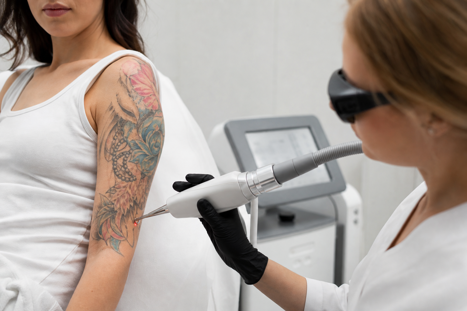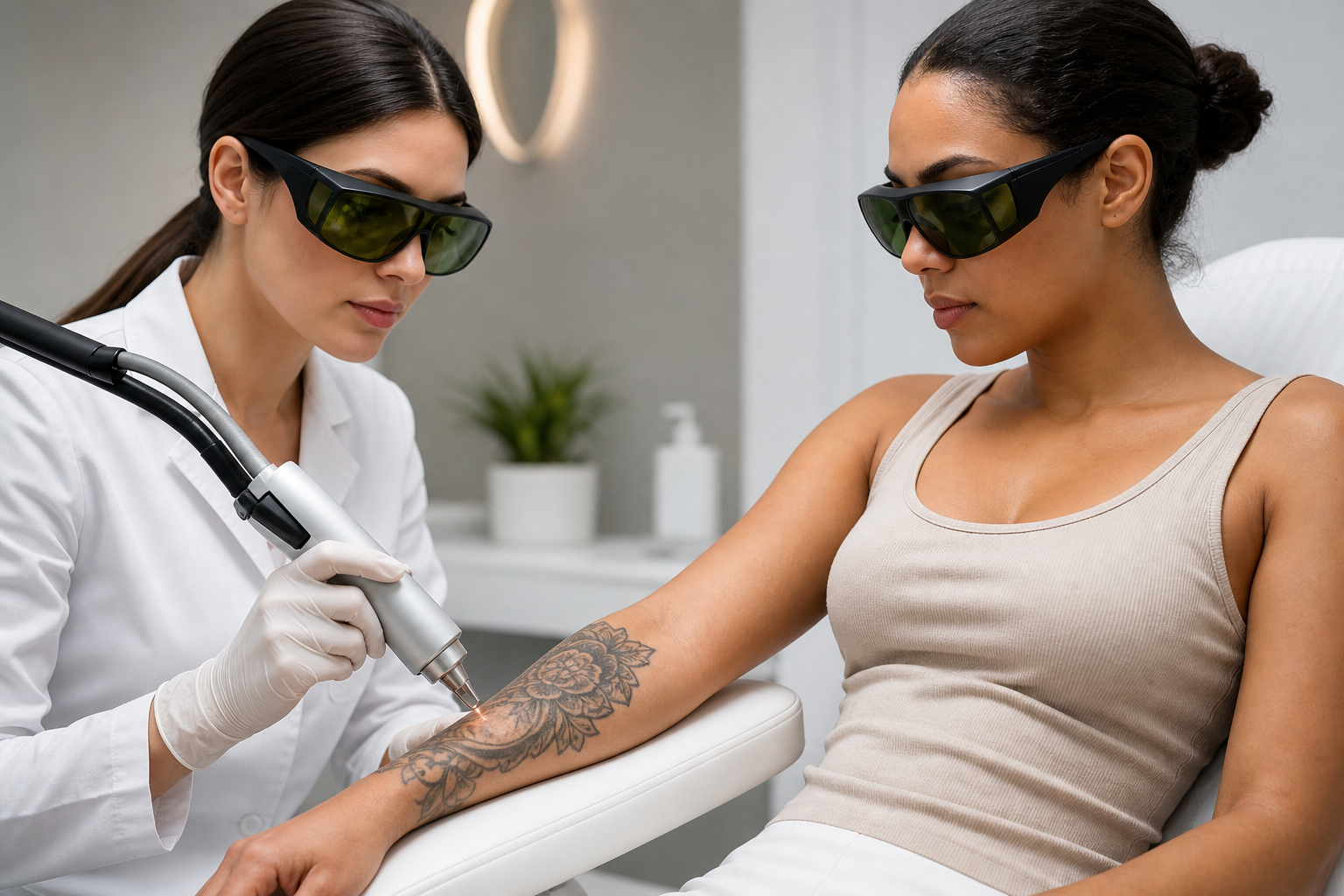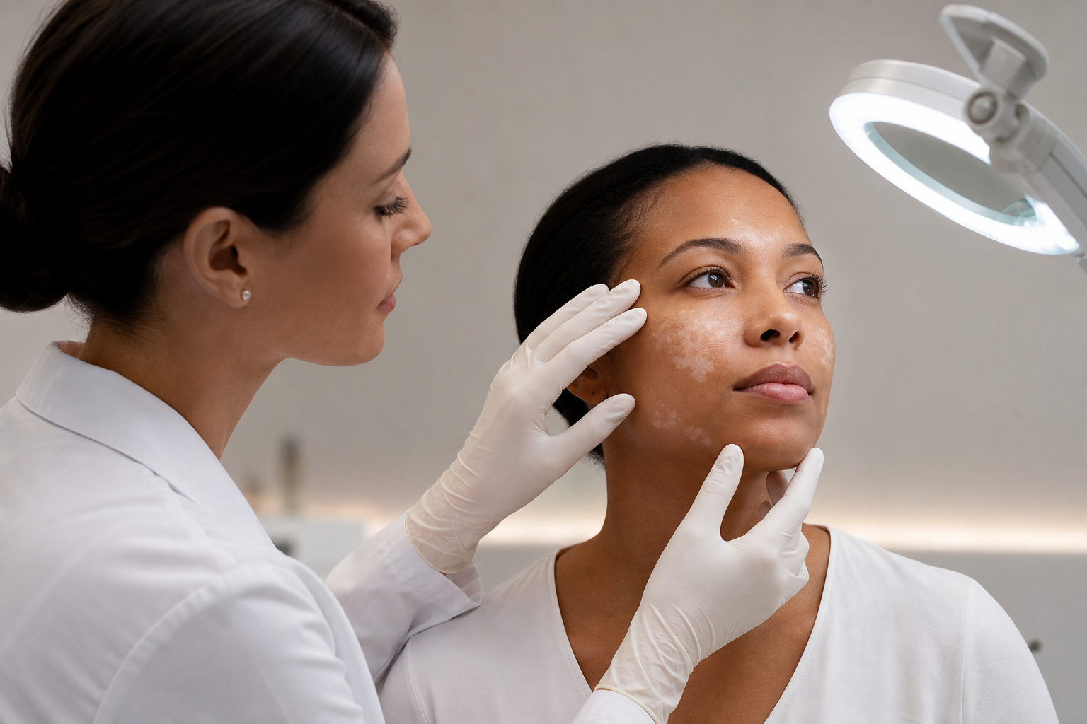
Hypopigmentation after laser treatment is a significant concern within dermatology and aesthetic medicine, characterized by lighter patches or depigmentation occurring in areas exposed to laser therapy. This phenomenon poses both aesthetic and psychological challenges for patients, particularly those with darker or tanned skin types. Understanding why hypopigmentation happens, which patient and device factors influence its development, and strategies for prevention and timely management is essential for anyone considering laser procedures or performing them.
This comprehensive article aims to provide an in-depth yet accessible exploration of post-laser hypopigmentation, addressing the needs of patients seeking clarity and clinicians striving to optimize treatment protocols while minimizing risks. As laser interventions become increasingly popular for skin rejuvenation and pigmentation correction, awareness of potential adverse effects such as hypopigmentation deepens the dialogue around safety and efficacy. Particularly for individuals with higher Fitzpatrick skin phototypes, appreciating the nuances of pigmentary changes is critical.
By examining the underlying causes, recognizing risk factors linked to individual skin characteristics and laser device parameters, and highlighting indicators that merit clinical review, this article contributes valuable educational insights. Readers should note, however, this information is not a substitute for personalized medical assessment and consultation with qualified healthcare professionals.
What Is Post-Laser Hypopigmentation and How Does It Present?
Hypopigmentation refers to a reduction in melanin production or distribution within the skin, resulting in areas that appear lighter than the surrounding tissue. After laser treatments—particularly those targeting pigmentation removal and skin resurfacing—patients may notice pale or depigmented spots at the treated sites.
This condition contrasts with the more commonly discussed post-inflammatory hyperpigmentation, where increased pigment deposition causes darkened patches. Hypopigmentation can present variably, ranging from subtle lightening to conspicuous white spots. The extent and duration may differ per individual; for some, these changes are transient, but for others, they can become persistent or permanent, complicating the cosmetic outcome.
Clinically, hypopigmentation manifests as well-demarcated, depigmented macules or patches. Texture changes such as smoothness or slightly roughened surfaces may accompany these areas. The visibility of these changes often depends on the baseline skin tone and the contrast created with the unaffected skin.
Which Patient Factors Influence the Risk of Hypopigmentation?
Diverse patient-related characteristics impact susceptibility to hypopigmentation following laser therapy. Recognizing these factors is foundational for informed treatment planning and patient counseling.
Skin Phototype: The Fitzpatrick scale categorizes skin types based on their response to ultraviolet (UV) exposure, ranging from I (very fair) to VI (very dark). Individuals with phototypes IV to VI generally possess higher melanin content, increasing the energy absorption during laser treatment. This heightened absorption predisposes melanocytes—the pigment-producing cells in the skin—to thermal damage, heightening the risk of post-treatment hypopigmentation.
Pre-existing Skin Conditions and Inflammation: Skin that has recently experienced inflammation, injury, or other dermatological conditions is more prone to pigmentary alterations. Such histories suggest a sensitized state, wherein melanocyte function or survival may already be compromised, increasing vulnerability to laser-induced pigment loss.
Sun Exposure: Acute ultraviolet exposure prior to or following laser therapy can exacerbate pigmentary instability. Tanned or sun-exposed skin contains more active melanocytes, which may respond unpredictably to laser energy and increase the risk for pigment disruption.
Genetic and Individual Susceptibility: Genetic predisposition influences the melanocyte's ability to regenerate after injury. Some individuals' melanocytes recover efficiently, restoring pigment over time, whereas others have a limited regenerative capacity, leading to longer-lasting or permanent hypopigmentation.
How Do Laser Device Characteristics Affect the Risk of Hypopigmentation?
The parameters and technology of laser devices play a crucial role in determining post-treatment pigmentary outcomes. Understanding these elements helps prevent adverse effects and enhance treatment safety.
Wavelength Selection: Different lasers operate at specific wavelengths targeting distinct chromophores, such as melanin or water. Selecting a wavelength suitable for the patient’s pigment type and treatment goals is vital. Incorrect wavelength selection can cause excessive damage to melanocytes, especially when treating pigmentation disorders.
Energy Levels and Fluence: High fluence or energy density can cause deeper thermal damage to the skin layers, increasing melanocyte destruction. Overly aggressive energy settings correlate with increased hypopigmentation risk.
Pulse Duration and Spot Size: Pulse duration affects how laser energy is delivered, with shorter pulses reducing collateral damage. Similarly, spot size influences energy distribution and penetration. Optimizing these parameters ensures targeted treatment while preserving surrounding tissues.
Device Technology and Features: Modern lasers often incorporate cooling systems, adjustable settings, and real-time feedback to minimize thermal injury. However, inappropriate use, lack of proficiency, or absence of a laser test spot assessment can elevate risk. Performing test spot treatments helps predict skin response and tailor parameters.
Why Are Caution and Tailored Approaches Essential for Darker and Tanned Skin?
Darker skin types and sun-exposed (tanned) skin present unique challenges during laser treatments. The high melanin content acts as a natural chromophore absorbing laser energy, which increases the chance of adverse pigmentary changes.
In darker skin, melanocytes are more active and abundant, making them particularly susceptible to laser-induced injury. Moreover, tanned skin is in an activated metabolic state with heightened melanocyte activity, rendering it more vulnerable to thermal damage. Consequently, clinicians must employ conservative energy settings, choose appropriate laser types, and carefully schedule treatments following adequate sun avoidance.
References such as "Fitzpatrick Skin Type and Laser Selection for Pigmentation" and "Pigmentation Lasers for Darker Skin" underscore these considerations and offer guidance on selecting safe and effective protocols. Further, the article "Why Does Tanned Skin Increase Treatment Risk?" elaborates on the pathophysiological basis for these heightened vulnerabilities.
What Precautions Can Minimize Hypopigmentation Before and After Laser Therapy?
Preventing hypopigmentation requires an integrated approach encompassing patient evaluation, laser parameter selection, and post-treatment care.
Comprehensive Patient Assessment: Prior to treatment, a thorough evaluation of skin phototype, history of pigmentary disorders, sun exposure, and previous skin damage is essential. Using the Fitzpatrick classification helps guide laser choice and settings. Medical history should include inquiries about autoimmune conditions or medications affecting pigmentation.
Performing Laser Test Spots: Conducting test spots on a small area allows clinicians to observe skin response, refining parameters and reducing the risk of unexpected pigmentary changes. The article "Laser Test Spots: When Are They Especially Important?" details best practices in this area.
Sun Avoidance: Preventing UV exposure days or weeks before and after treatment stabilizes melanocyte activity, lowering pigment-related complications. Patients should use broad-spectrum sunscreens, wear protective clothing, and avoid tanning.
Customized Laser Settings: Tailoring energy, wavelength, pulse duration, and treatment density to the individual's skin features ensures efficient therapy with minimal collateral damage.
Post-Treatment Skin Care: Application of calming, regenerative topical agents under medical guidance supports skin healing and pigment recovery. Continuous monitoring helps detect early signs of adverse pigmentary responses.
Which Symptoms Should Prompt Immediate Clinical Evaluation?
Early recognition of hypopigmentation signs enables timely intervention, potentially mitigating progression or permanence.
Emergence of Light or White Patches: Noticeable color changes not present before treatment require attention, especially if sharply demarcated and persistent.
Altered Skin Texture: Accompanying changes such as smoothness, dryness, or roughness at the treated site may indicate melanocyte or epidermal disruption.
Unusual Sensations: Persistent cooling sensations, numbness, or unusual skin sensitivity could signal deeper tissue injury associated with altered pigmentary function.
Any of these developments merit consultation with a dermatologist or trained laser specialist to evaluate severity, review treatment strategy, and guide corrective measures.
How Is Laser-Induced Hypopigmentation Treated and Managed?
Addressing hypopigmentation requires a multifaceted approach centered on stimulating melanocyte recovery and modulating skin pigmentation.
Topical Therapies: Prescription retinoids can enhance epidermal turnover and promote melanocyte activation. Other topical agents such as corticosteroids may reduce inflammation that could impede pigment restoration.
Phototherapy: Controlled ultraviolet light exposure under physician supervision may stimulate melanocyte proliferation and melanin synthesis.
Microneedling: This minimally invasive procedure induces skin regeneration pathways and may normalize pigmentation patterns by promoting melanocyte migration.
Careful Subsequent Laser Treatments: In select cases, gentle laser therapy with adjusted parameters can even out skin tone but must be approached cautiously due to inherent risks.
Time is a considerable factor, as pigment recovery may extend over several months. Patients should maintain realistic expectations and engage in close follow-up with their healthcare providers.
What Common Patient Concerns Arise and How Can They Be Addressed?
Patients often express anxiety about the permanence of hypopigmentation and seek clarity on treatment outcomes. Providing transparent communication is foundational.
Emphasizing Prevention: Clarifying that individualized assessment and cautious protocol tailoring decrease adverse effects mitigates undue worry.
Distinguishing Different Reactions: Educating patients on the difference between typical transient pigment fluctuations and more serious hypopigmentation fosters informed vigilance.
Presenting Treatment Options: Outlining correction modalities, limitations, and anticipated timeframes helps patients set achievable goals.
Encouraging Open Dialogue: Ensuring patients feel comfortable reporting changes promptly supports proactive management.