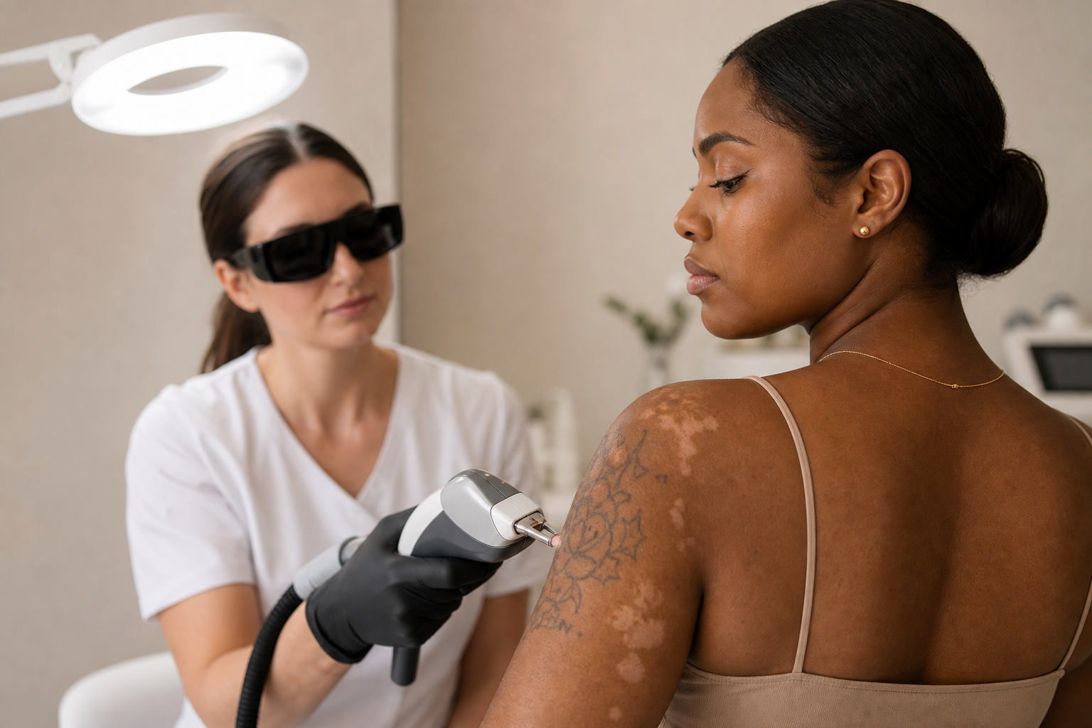
In the realm of dermatology and cosmetic treatments, understanding the nuances of skin pigmentation is crucial. Hyperpigmentation and hypopigmentation are two contrasting conditions that affect the coloration of skin, resulting in either darker or lighter patches relative to the surrounding areas. These pigmentary changes arise due to alterations in melanin synthesis or distribution, and they have significant implications for both patients and practitioners, especially when involving advanced procedures such as laser tattoo removal.
This article delves deeply into the distinctions between hyperpigmentation and hypopigmentation, exploring their causes, clinical presentations, and relevance to individuals seeking cosmetic or therapeutic skin interventions. It also highlights the potential risks associated with laser treatments, particularly in individuals with darker or tanned skin, emphasizing the need for professional assessment and carefully tailored treatment protocols.
By providing comprehensive insights into these pigmentary phenomena, this editorial aims to inform patients considering laser tattoo removal and other pigmentation-related therapies, as well as medical professionals who perform these procedures. Understanding the underlying mechanisms and risks is essential to optimize treatment outcomes and minimize adverse effects.
Defining Hyperpigmentation: Causes and Common Manifestations
Hyperpigmentation is characterized by localized or diffuse darkening of the skin due to an excess production or accumulation of melanin. Melanin, the natural pigment responsible for the color of skin, hair, and eyes, serves as a protective shield against ultraviolet (UV) radiation. However, when melanocytes—the cells that produce melanin—become overactive or unevenly stimulated, it leads to irregular coloration patterns on the skin.
Common forms of hyperpigmentation include melasma, solar lentigines (often referred to as age spots or liver spots), and post-inflammatory hyperpigmentation (PIH). Melasma typically presents as symmetric brown patches and is more prevalent among women, particularly during hormonal fluctuations such as pregnancy or with the use of oral contraceptives. Solar lentigines are caused by chronic sun exposure, resulting in well-demarcated pigmented spots usually found on sun-exposed areas like the face and hands.
Post-inflammatory hyperpigmentation arises following skin injury or inflammation, such as after acne, eczema, or trauma. These darker marks represent the skin’s healing response but can persist for extended periods, posing aesthetic concerns.
Although hyperpigmentation is generally benign, it can profoundly impact an individual’s self-esteem, motivating many to pursue treatment options ranging from topical agents to advanced laser therapies.
Understanding Hypopigmentation and Its Clinical Implications
In contrast to hyperpigmentation, hypopigmentation denotes a reduction or complete absence of melanin in the affected skin areas, leading to lighter or nearly white patches. This shift in pigmentation can be congenital or acquired.
Vitiligo stands as the quintessential example of congenital hypopigmentation, where an autoimmune process targets and destroys melanocytes, resulting in well-defined depigmented areas scattered symmetrically or irregularly across the body. Unlike hyperpigmentation, these patches can rapidly expand and often require specialized management.
Acquired hypopigmentation may result from trauma, burns, infections, or as unintended consequences of medical procedures like laser treatments. When lasers are improperly used—whether due to excessive intensity, incorrect wavelength, or inadequate technique—they can damage melanocytes, leading to partial or total pigment loss in the targeted skin regions.
Distinguishing hypopigmentation from scarring or other textural changes is critical for an accurate diagnosis and appropriate therapeutic planning.
Who Should Understand the Differences Between Hyperpigmentation and Hypopigmentation?
Recognizing these pigmentary conditions is vital for a broad spectrum of individuals. Patients experiencing noticeable changes in skin color—whether darkened or lightened spots—must comprehend the nature of their condition to make informed decisions about seeking treatment.
Moreover, dermatologists, laser technicians, and practitioners specializing in aesthetic medicine need to have a robust understanding of pigmentary disorders. This knowledge equips them to select suitable treatment modalities, customize parameters according to individual skin types, and anticipate possible complications, ensuring safety and satisfaction.
The importance of this understanding amplifies in populations with darker or tanned skin, where the balance of melanin is inherently different, and the risk of post-treatment pigmentary alterations rises significantly. Such patients require careful pre-treatment evaluation and post-care to minimize adverse pigmentary outcomes.
Mechanisms Behind Pigment Changes Following Laser Tattoo Removal
Laser tattoo removal employs focused light energy to disrupt tattoo ink particles embedded within the dermis. The photoacoustic effect breaks down these ink clusters into smaller fragments that the body’s immune system can gradually clear. This technique is considered highly effective and less invasive than surgical removal.
Despite its advantages, laser tattoo removal poses risks of pigmentary disturbances. Hyperpigmentation can develop when the skin responds to laser-induced inflammation with increased melanocyte activity, producing excess melanin in the treated region. This response is particularly pronounced if sun protection is inadequate post-procedure, as ultraviolet exposure further stimulates melanin synthesis.
Conversely, hypopigmentation occurs when laser energy damages or destroys melanocytes. This injury may be irreversible, leading to persistent pale patches contrasting with the normal skin tone. Individuals with naturally higher melanin levels or recent tanning are more predisposed to this effect, necessitating adapted laser settings and meticulous treatment planning.
Understanding these mechanisms helps both clinicians and patients set realistic expectations and adopt preventive strategies to mitigate risks.
Crucial Risk Factors and Prevention Strategies for Pigmentation Disorders Post-Laser Treatment
Several factors influence the likelihood of developing hyper- or hypopigmentation after laser tattoo removal or similar interventions. Key determinants include the individual's Fitzpatrick skin phototype, the depth and color composition of the tattoo pigment, laser parameters (wavelength, pulse duration, and fluence), and the practitioner's expertise.
Darker skin types possess more active melanocytes and higher melanin content, increasing susceptibility to pigment changes. Inappropriate laser settings or over-treatment may easily damage melanocytes, leading to hypopigmentation, whereas under-treatment or inflammatory responses can result in hyperpigmentation.
Sun exposure surrounding the treatment period elevates the risk dramatically. UV radiation activates melanocytes, worsening pigmentary irregularities, hence stringent photoprotection using broad-spectrum sunscreens and physical barriers is indispensable.
Professionalism in treatment execution is equally important. Experienced dermatologists and laser specialists contribute to safer outcomes through precise diagnosis, selecting appropriate laser systems tailored to the patient’s skin type and tattoo characteristics, and timely monitoring and managing adverse effects.
Importantly, patients should avoid unverified home remedies or do-it-yourself approaches that may aggravate skin damage or pigmentation abnormalities.
Common Skin Reactions After Laser Procedures and When to Seek Dermatological Advice
Following laser tattoo removal and pigmentation treatments, some degree of skin reaction is expected. Immediate responses usually include mild redness, swelling, serous exudate, and crusting, which generally resolve within a week. These are considered normal phases of healing.
However, persistent inflammation, intense pain, ulceration, signs of infection, or expanding pigmentary changes warrant prompt professional evaluation. The emergence of new hyperpigmented or hypopigmented spots within several weeks post-treatment should alert both patient and practitioner to possible complications requiring intervention.
Pre-treatment consultation is advisable, especially for individuals with darker or recently tanned skin, to establish baseline skin condition, discuss risks, and customize treatment protocols accordingly.
The Importance of Dermatologist Evaluation Before Laser Tattoo Removal
Before initiating laser tattoo removal, a thorough assessment by a qualified dermatologist is essential. This examination includes evaluating the tattoo’s ink type, depth, and color, as well as the skin's overall health and condition surrounding the tattoo.
A dermatologist can identify contraindications such as active skin infections, inflammatory conditions, or scarring tendencies, which may affect treatment safety. Additionally, in darker-skinned patients, preemptive strategies to prevent pigmentary complications can be implemented.
This preliminary step not only facilitates informed consent but also contributes to developing an individualized treatment plan that maximizes efficacy and minimizes adverse effects like hyperpigmentation or hypopigmentation.