Post-inflammatory hyperpigmentation (PIH) manifests as darkened patches or spots on the skin following inflammatory processes such as acne breakouts, eczema, or other dermatological irritations. For many individuals, these dark spots become a stubborn cosmetic concern, detrimentally affecting self-esteem and complicating skincare routines. The emergence of advanced laser technologies, particularly the picosecond laser, has ushered in new possibilities for effectively addressing these challenging pigmentary issues. However, understanding the nuances between different post-inflammatory discolorations, identifying suitable candidates, and appreciating the risks involved are crucial steps before engaging in laser therapy.
This article delves into the role of picosecond laser treatment for post-inflammatory dark spots, elaborating on its mechanism, suitability for diverse skin types, benefits, limitations, and precautions. Dermatologists, skincare professionals, and individuals grappling with post-inflammatory pigmentation will find comprehensive insights to guide informed decisions on this sophisticated treatment modality.
Navigating the complexities of pigment correction requires distinguishing between the brown hues of true post-inflammatory hyperpigmentation and red post-inflammatory erythema, two conditions arising from separate pathological processes. This distinction underlines the importance of targeted therapy to avoid exacerbation or suboptimal outcomes. Furthermore, special caution is warranted for patients with darker skin phototypes, due to the heightened risk of paradoxical pigmentation changes after laser exposure.
What Is the Picosecond Laser and How Does It Address Post-Inflammatory Dark Spots?
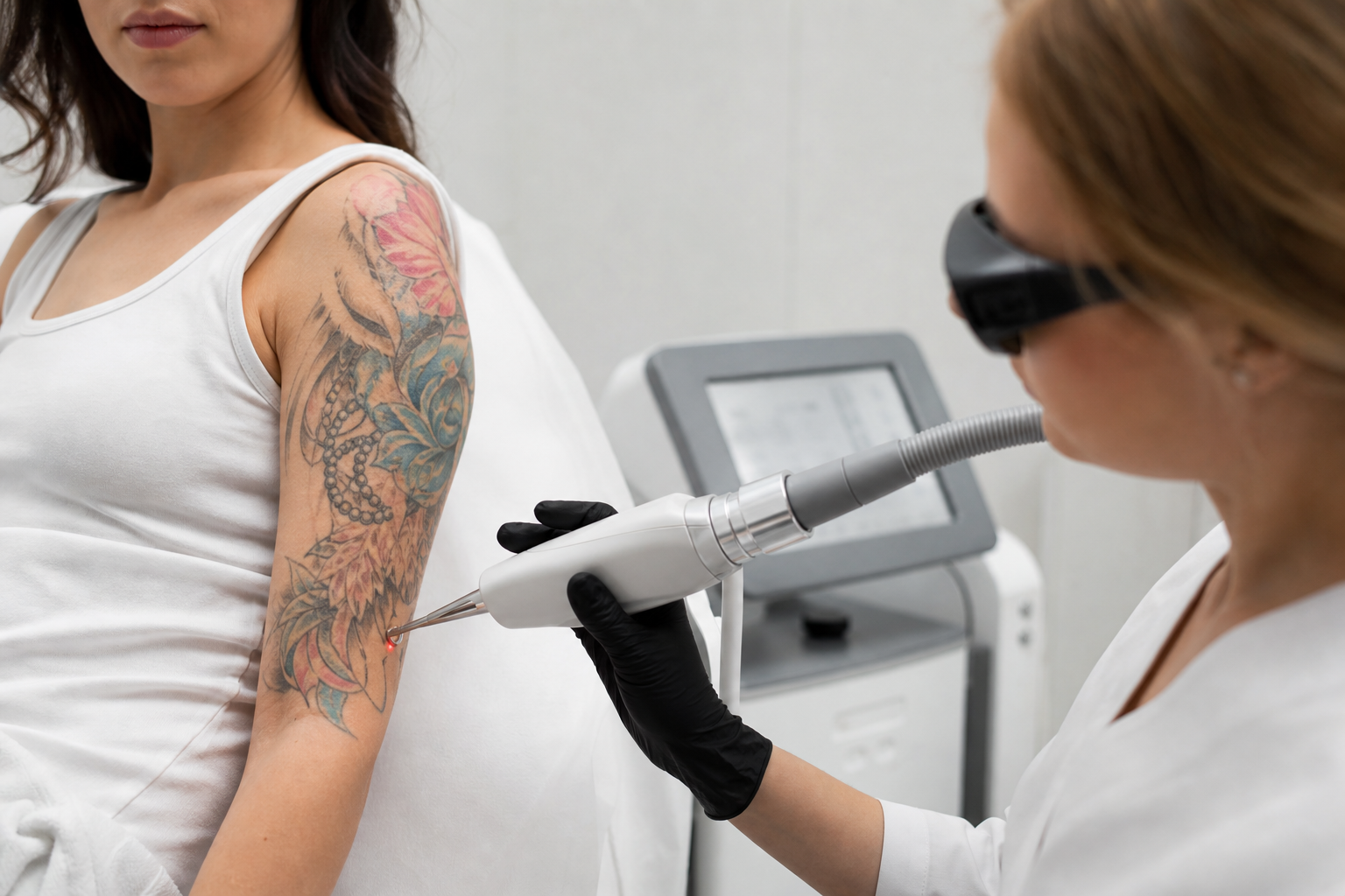
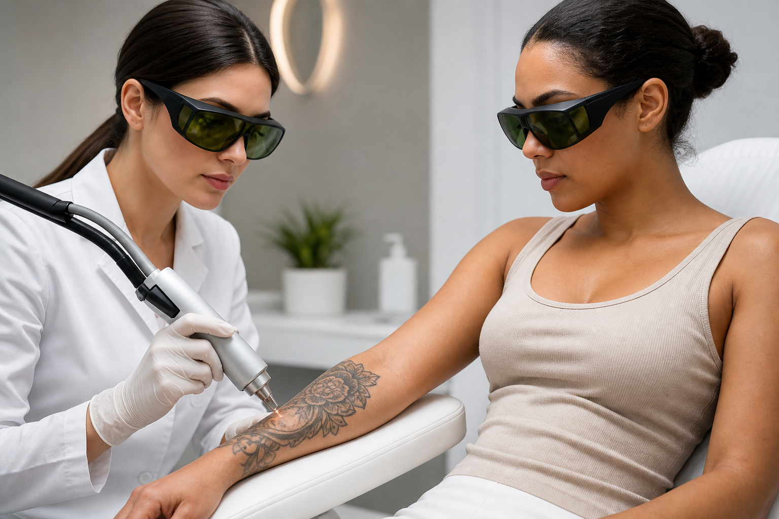
The picosecond laser represents an innovative class of laser devices that deliver ultrashort light pulses measured in picoseconds (one trillionth of a second). This extremely brief pulse duration enables the delivery of high energy bursts to pigment-laden cells, resulting in their mechanical fragmentation rather than heat-mediated damage. Consequently, picosecond lasers can selectively target melanin deposits within the skin with minimal collateral tissue injury.
In cases of post-inflammatory hyperpigmentation, melanin accumulates unevenly due to an overproduction triggered by prior skin inflammation. The pigment exists across varying depths within the epidermis and dermis, necessitating a laser modality capable of precise penetrative action. Picosecond lasers employ photomechanical effects to shatter melanin granules into minuscule particles, which are then naturally cleared by macrophages and lymphatic drainage, leading to gradual lightening of dark spots.
Contrast this with traditional nanosecond lasers, which rely more heavily on photothermal effects that can generate higher heat and potentially induce unwanted skin injury or inflammation. The picosecond mechanism enhances safety and efficacy, making it particularly apt for intricate pigmentation abnormalities where preservation of surrounding skin integrity is paramount.
Distinguishing Brown Post-Inflammatory Hyperpigmentation from Red Post-Inflammatory Erythema
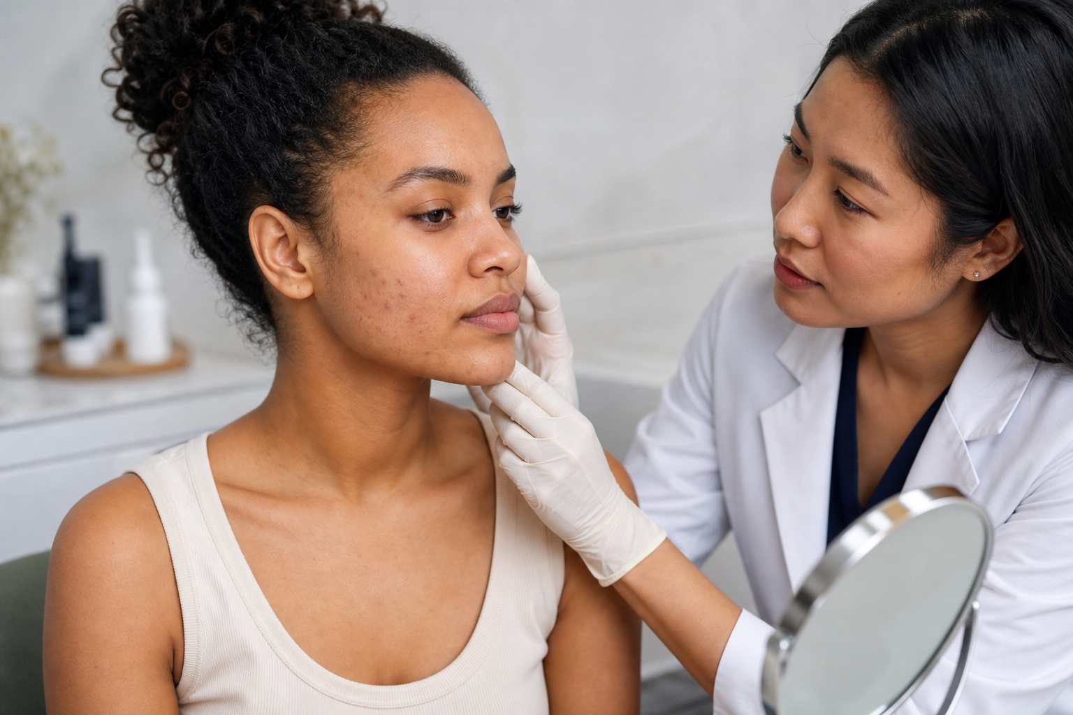
One often overlooked but critical diagnostic facet in pigment treatment involves differentiating between brown PIH and red post-inflammatory erythema. Brown PIH arises principally from melanin deposition triggered by skin injury or inflammation, resulting in hyperpigmented areas usually darker than the surrounding skin. These spots tend to persist long after the initial insult subsides and are responsive to pigment-targeting interventions such as picosecond lasers.
Conversely, red post-inflammatory erythema stems from vascular changes, specifically prolonged dilation or damage to superficial blood vessels following inflammation. This manifests as reddish or pink areas which are not true pigmentation changes but vascular discolorations. Treating these with pigment-targeting lasers like picosecond devices may prove ineffective or even detrimental, potentially aggravating erythema or causing irritation. Instead, vascular lasers or other specialized therapies are typically more appropriate.
The clinical implication is clear: accurate diagnosis through visual assessment, dermoscopy, and sometimes adjunctive imaging or biopsy is essential to selecting the correct treatment path. Failure to distinguish these entities can result in therapeutic failure or paradoxically worsening discoloration.
Why Controlling Active Inflammation is Crucial Before Picosecond Laser Treatment
Active skin inflammation is a contraindication for laser interventions targeting post-inflammatory dark spots. Engaging laser treatment while the skin is still inflamed risks exacerbating irritation, prolonging healing time, and provoking additional pigmentation changes or scarring. Before initiating laser therapy, all instances of active acne, eczema flares, or dermatitis should be thoroughly managed and stabilized.
Managing active inflammation may encompass topical anti-inflammatory agents, systemic medications when appropriate, and lifestyle or environmental modifications that mitigate flare triggers. Only once the inflammatory activity has subsided and the skin surface has normalized is it safe to proceed with the picosecond laser to address residual hyperpigmentation.
This preparatory phase not only safeguards skin health but also optimizes treatment outcomes, as the laser-induced pigment clearance acts on stable pigment deposits rather than transient erythematous changes.
Special Considerations for Treating Darker Skin Phototypes with Picosecond Lasers
Darker skin phototypes (Fitzpatrick IV-VI) present unique challenges and risks in laser therapy due to their increased baseline melanin content, which heightens susceptibility to post-inflammatory hyperpigmentation or hypopigmentation following skin trauma. Though picosecond lasers' precision and low thermal damage profile make them comparatively safer, meticulous parameter adjustments and clinical judgment remain vital.
Practitioners must carefully titrate fluence, pulse frequency, and spot size to avoid overt epidermal insult, while also incorporating thorough patient education regarding sun protection, skincare maintenance, and symptom monitoring. Regular follow-up enables prompt detection and management of any adverse pigmentary responses.
Equally important is adopting a personalized approach without relying on universal protocols; treatment plans should be customized based on skin type, pigmentation depth, patient history, and concurrent skin conditions to minimize the risk of laser-induced darkening or other complications.
Potential Risks and Adverse Effects: Can Picosecond Laser Treatment Worsen Pigmentation?
While picosecond laser therapy is regarded as a leading-edge approach for treating pigmentary disorders, it is not devoid of risks. One potential adverse effect includes paradoxical hyperpigmentation, whereby the treatment intended to ameliorate dark spots inadvertently stimulates melanocyte activity, leading to darkening of the area. Such rebound pigmentation may arise from improper treatment timing, inadequate control of active inflammation, excessive laser energy settings, or insufficient photoprotection post-procedure.
Furthermore, inadvertent injury to the skin barrier during treatment can result in irritation, inflammation, or erythema, potentially precipitating new pigmentary changes. These risks are amplified in patients with darker phototypes or a history of pigmentary disorders.
Mitigation strategies include conservative treatment parameters, thorough patient screening, rigorous sun avoidance practices following therapy, and use of topical agents to support skin barrier repair and pigment modulation. Educating patients to set realistic expectations and adhere to post-treatment care protocols is equally essential to achieving favorable safety and efficacy.
Timing the Treatment: When Should Picosecond Laser Therapy for Post-Inflammatory Dark Spots Be Initiated?
Determining the optimal timing for picosecond laser intervention is pivotal to maximizing benefits and minimizing complications. Generally, a period of quiescence following the active inflammatory episode is advised—this often translates to several weeks or months of skin recovery to allow inflammation to fully subside and early pigmentation changes to stabilize.
Launching treatment prematurely during the acute phase may fail to achieve lasting pigment clearance and elevate the risk of irritation or adverse pigmentary responses. Diagnostic assessments, including photographic documentation and clinical examination, assist in appraising lesion maturity and readiness for laser therapy.
Patient-specific factors such as extent of pigmentation, skin type, history of laser treatments, and concurrent skin conditions also influence timing decisions. Accordingly, individualized treatment planning is paramount to align laser therapy with dermatological best practices and patient needs.
What Can Patients Expect During and After Picosecond Laser Treatment Sessions?
Picosecond laser sessions typically last from a few minutes up to half an hour depending on the size and number of treated areas. Patients often experience mild sensations described as tingling or snapping during laser application, which is usually well-tolerated with optional topical anesthetics or cooling devices enhancing comfort.
Immediately post-treatment, transient redness, mild swelling, or a sensation of skin tightness can occur, typically resolving within several days. Some patients may notice slight dryness or flaking as the skin renews itself. Crucially, dark spots do not vanish instantly but lighten progressively as the body clears fragmented pigment over weeks.
Completion of multiple treatment sessions, spaced by approximately four to six weeks, is common to achieve optimal results. Post-procedural care encompasses diligent hydration, sun protection using broad-spectrum high-SPF sunscreens, and adherence to any prescribed topical agents. Regular follow-up visits facilitate monitoring of epidermal healing and pigment resolution.
Common Questions About Picosecond Laser for Post-Inflammatory Hyperpigmentation
Is picosecond laser suitable for all skin types? While effective across a broad range of skin phototypes, darker skin tones require specialized protocols and experienced practitioners to minimize pigmentation risks.
How many sessions are generally needed? The treatment course varies individually but commonly ranges from three to six sessions.
Can it be combined with other treatments? Yes, with careful planning, picosecond laser can be integrated with topical agents, chemical peels, or other aesthetic procedures to enhance outcomes.
What protective measures should be followed after treatment? Consistent use of sunscreen, avoidance of direct sunlight, and adherence to dermatologist-recommended skincare regimens substantially support healing and prevent complications.