Pigmented lesions on the skin encompass a broad spectrum of discolorations that can arise from various causes such as sun exposure, age, hormonal changes, or inflammatory processes. Managing these lesions effectively requires a precise and scientifically grounded approach, especially when considering laser therapies. Among the advanced laser technologies, the Alexandrite laser stands out for its ability to target melanin-rich skin abnormalities with a specific wavelength and mechanism tailored to pigment removal. This article delves into the nuances of Alexandrite laser treatment for pigmented lesions, providing a detailed understanding for patients exploring options and clinicians aiming to refine their therapeutic arsenal.
Understanding this technology is vital, not only because pigmented lesions can significantly affect a person's aesthetic appearance and self-esteem but also because the choice of laser must align with an individual's skin type and lesion characteristics to ensure safety and effectiveness. The Alexandrite laser offers promising outcomes for selected cases, but like any medical treatment, it carries limitations and requires expert handling to maximize benefits and minimize risks.
This comprehensive editorial serves those seeking in-depth knowledge about how the Alexandrite laser functions in pigment removal, including its mechanisms, potential indications, limitations, patient selection criteria, safety concerns, and how it compares within the broader field of laser therapies for pigmentation. By grounding the discussion in current dermatologic practice standards, it aims to inform intelligent decision-making, whether for personal treatment consideration or clinical application.
What Is the Alexandrite Laser and How Does It Work for Pigmented Lesions?
The Alexandrite laser is a solid-state laser utilizing a synthetic alexandrite crystal to produce a specific wavelength of light at approximately 755 nanometers, which lies within the near-infrared spectrum. This wavelength demonstrates a marked affinity for melanin, the pigment responsible for coloring skin, hair, and eyes. Selective absorption of the laser energy by melanin allows the Alexandrite laser to disrupt melanin-containing structures in pigmented lesions while sparing the surrounding non-pigmented tissue.
The core mechanism is selective photothermolysis. When pulsed laser light targets melanin, the pigment rapidly absorbs the light energy and converts it into heat. This localized thermal effect damages the pigment-containing cells, fragmenting excess melanin and pigmented cells without significantly affecting neighboring tissues. The body's natural immunologic and metabolic systems subsequently clear the damaged pigment and cellular debris, leading to a reduction or removal of the visible pigmented lesion.
This precise targeting makes the Alexandrite laser particularly suited for treating epidermal pigmented lesions such as lentigines, solar lentigo (sun spots), and certain post-inflammatory hyperpigmentations. Its depth of penetration and wavelength enable effective treatment of superficial lesions while maintaining a favorable safety profile for lighter skin types.
Which Pigmented Lesions Are Best Treated with the Alexandrite Laser?
The Alexandrite laser's efficacy is closely tied to the location and nature of pigmentation within the skin layers. It is best suited for epidermal pigmented lesions where excess melanin or melanocyte proliferation is superficial. Common indications include age spots or lentigines typically seen in sun-damaged skin, benign hyperpigmented areas resulting from inflammation (post-inflammatory hyperpigmentation), and some types of melasma, particularly when pigmentation is primarily epidermal.
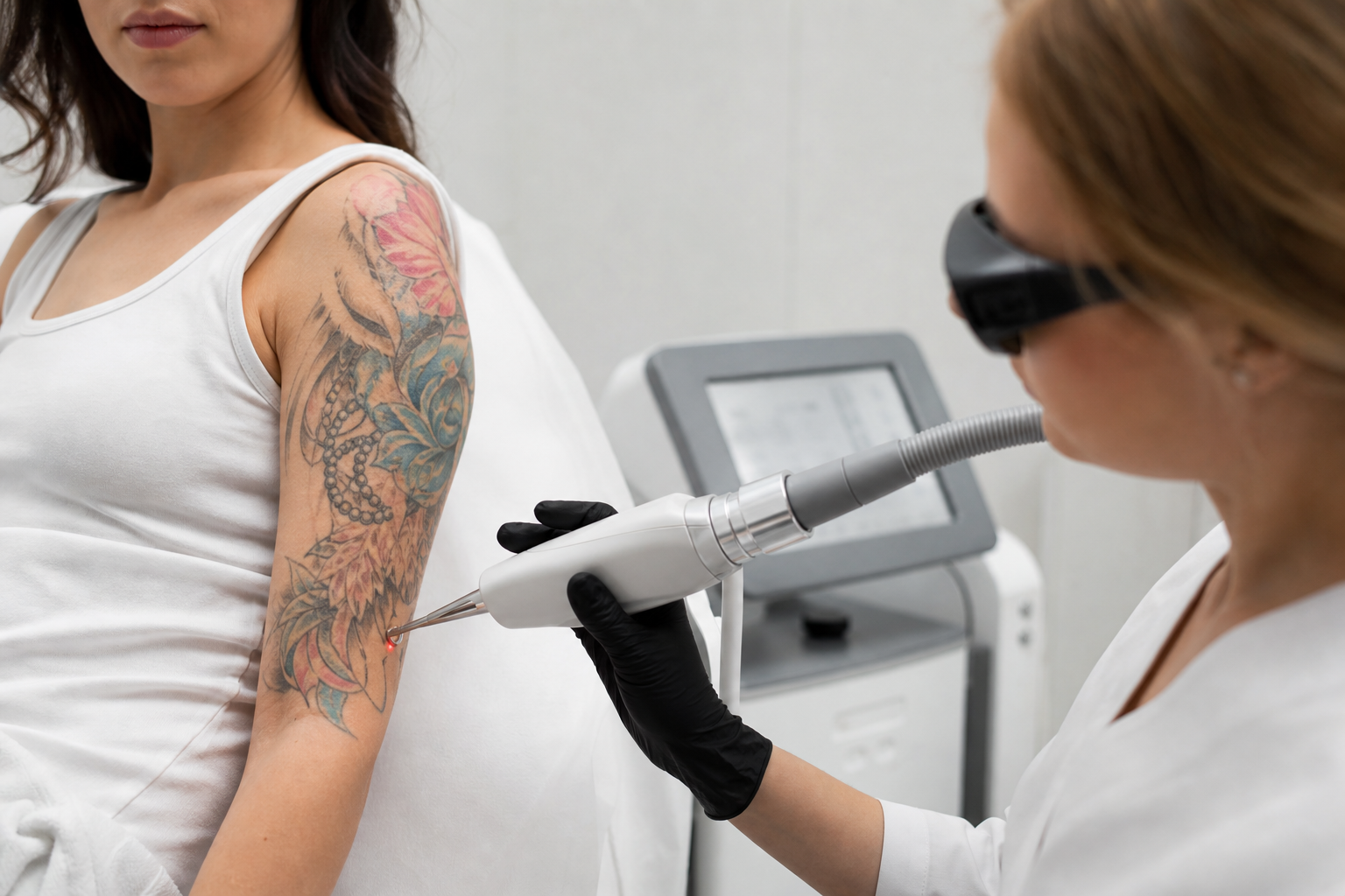
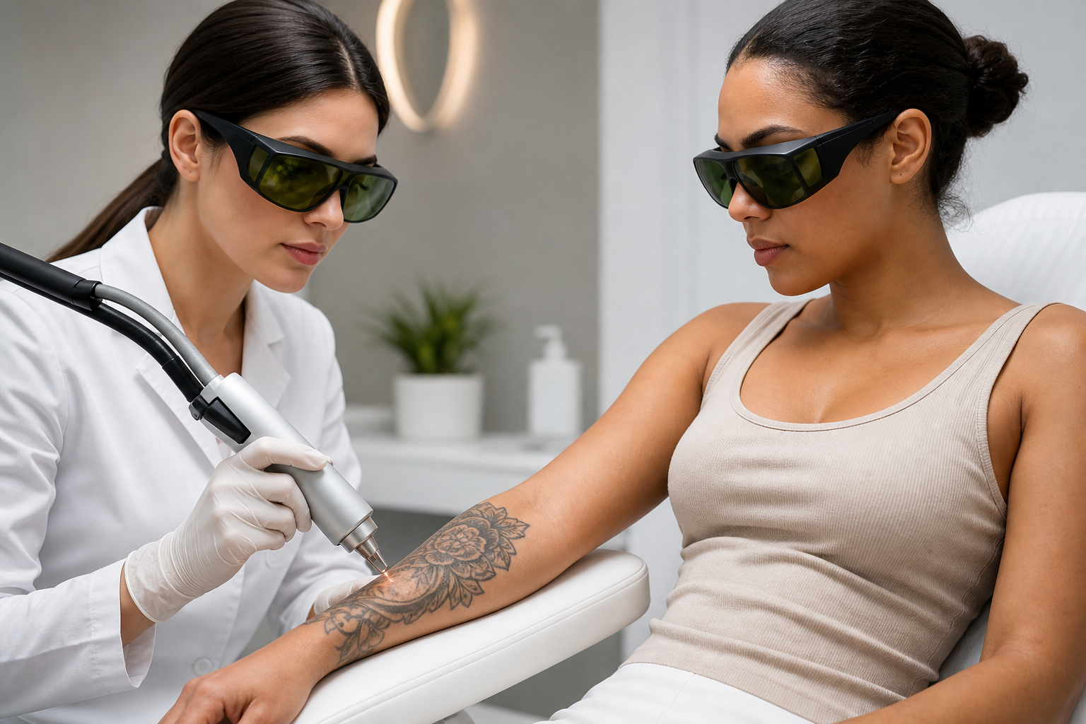
The laser is also utilized for the removal of certain dark tattoos due to its melanin-targeted mechanism and has a role in permanent hair reduction by targeting melanin in hair follicles, especially in patients with lighter skin phototypes. However, when specifically addressing pigmented lesions, its advantages are significant for patients with Fitzpatrick skin types I through III, where the contrast between lesion pigmentation and surrounding skin allows safer and more effective energy delivery.
Lesions with clear borders and those not deeply embedded within dermal layers respond better to Alexandrite laser treatments. Conditions such as solar lentigines, ephelides (freckles), and some types of benign congenital nevi may be amenable to this laser under proper clinical assessment.
What Are the Limitations of the Alexandrite Laser in Treating Pigmented Lesions?
Despite its benefits, the Alexandrite laser is not universally effective for all pigmented lesions. One of the main constraints is its diminished efficacy and increased risk profile in treating deeper dermal pigmentation or mixed epidermal-dermal pigmentation, such as certain presentations of melasma or nevus of Ota. These lesions may require alternative or adjunctive laser modalities with different wavelengths or delivery methods.
The laser's relatively high melanin absorption means that it can pose risks to patients with darker skin types (Fitzpatrick IV-VI), where melanin is more abundant in the surrounding skin, increasing the chance of adverse effects like hypopigmentation, hyperpigmentation, or scarring. Hence, treatments in these populations often necessitate lasers with wavelengths less absorbed by melanin, such as Nd:YAG lasers operating at 1064 nm or the use of picosecond pulse durations to minimize thermal damage.
Additionally, the laser's effectiveness is dependent on an accurate diagnosis of the pigmented lesion to exclude malignancy (e.g., melanoma), vascular lesions, or other skin conditions that may superficially mimic pigmentation. Misapplication can lead to inadequate results or exacerbation of skin issues.
How Is Skin Phototype Assessed, and Why Is It Crucial for Alexandrite Laser Treatment?
Precisely classifying a patient's skin phototype is fundamental in predicting skin response to laser treatment and adjusting parameters accordingly. The Fitzpatrick Skin Phototype scale, ranging from I (very fair skin, always burns, never tans) to VI (deeply pigmented dark skin, never burns), is commonly used to categorize skin sensitivity to ultraviolet light and melanin quantity.
Since the Alexandrite laser targets melanin selectively, skin types with less melanin (I to III) allow clearer differentiation between pigmented lesions and surrounding tissue. This distinction reduces the risk of unwanted laser energy absorption by non-lesional skin, lowering complication rates. Conversely, higher Fitzpatrick types require cautious parameter adjustments or alternative laser selection to avoid damage to the surrounding melanin-rich epidermis.
Clinicians employ additional diagnostic tools alongside visual examination, such as dermoscopy and sometimes spectrophotometry or confocal microscopy, to assess lesion depth, melanin concentration, and skin characteristics. This comprehensive evaluation informs the safest and most efficacious treatment plan tailored to the individual.
What Are the Safety Considerations and Potential Risks of Alexandrite Laser Treatment?
Although Alexandrite laser treatment is generally safe when performed by qualified professionals, it carries intrinsic risks primarily due to its selective targeting of melanin-rich areas. Common side effects include transient erythema (redness), edema (swelling), and mild discomfort immediately following treatment. These effects typically resolve within hours to a few days.
More serious, albeit less frequent, complications encompass blistering, crusting, hyperpigmentation or hypopigmentation, and in rare cases, scarring. These are largely preventable through correct patient selection, accurate phototype assessment, meticulous laser parameter customization, and strict adherence to pre- and post-treatment care guidelines.
Pre-treatment precautions involve avoiding sun exposure and tanning for several weeks, as increased melanin can heighten the risk of adverse effects. The use of broad-spectrum sunscreens and sun-protective behaviors must continue post-procedure to maintain results and prevent recurrence or worsening of pigmentary changes.
Informed consent and comprehensive patient education regarding realistic expectations, potential risks, and the necessity for multiple sessions are essential components of the treatment process. Follow-up evaluations enable early detection and management of any complications, ensuring optimal healing and outcomes.
Which Device Parameters Influence the Alexandrite Laser's Effectiveness and Safety?
Therapeutic success with the Alexandrite laser hinges on the careful adjustment of key device parameters such as fluence (energy density), pulse duration, pulse repetition rate, and spot size. Fluence determines the amount of energy delivered per unit area; higher fluence enhances pigment destruction but raises the risk of collateral tissue injury.
Pulse duration must be aligned with the thermal relaxation time of targeted melanin granules to achieve selective photothermolysis without excessive heat dispersal. Shorter pulses allow targeted damage with minimal surrounding tissue effects, while longer pulses may increase safety but reduce efficacy on small pigment particles.
Spot size affects treatment area coverage and penetration depth. Larger spot sizes permit deeper light penetration and faster treatment but may reduce precision. Repetition rate influences treatment speed and skin cooling intervals, balancing efficacy with patient comfort and safety.
Experienced clinicians tailor these parameters individually, taking into account lesion type, depth, patient skin characteristics, and treatment goals. Protocols may evolve throughout a course of sessions based on response and tolerability.
Why Must a Qualified Professional Oversee Alexandrite Laser Treatment?
The complexity inherent in diagnosing pigmentary disorders, selecting suitable candidates, and tailoring laser parameters demands expertise found in dermatologists or laser-trained medical professionals. Misuse or lack of precise assessment risks serious adverse outcomes, from ineffective treatment to permanent skin damage.
Qualified practitioners are also equipped to distinguish benign pigmented lesions from suspicious or malignant ones, ensuring that laser therapy is appropriate and deferring to biopsy or oncology consultation when needed. Moreover, they can manage unexpected reactions, provide adjunctive care, and integrate laser therapy within a comprehensive skin treatment plan.
Ongoing education, hands-on training, and adherence to best practice guidelines form the foundation of delivering Alexandrite laser treatments safely and effectively. This oversight reassures patients and upholds the standards of dermatologic care.
What Can Patients Expect During and After Alexandrite Laser Treatment?
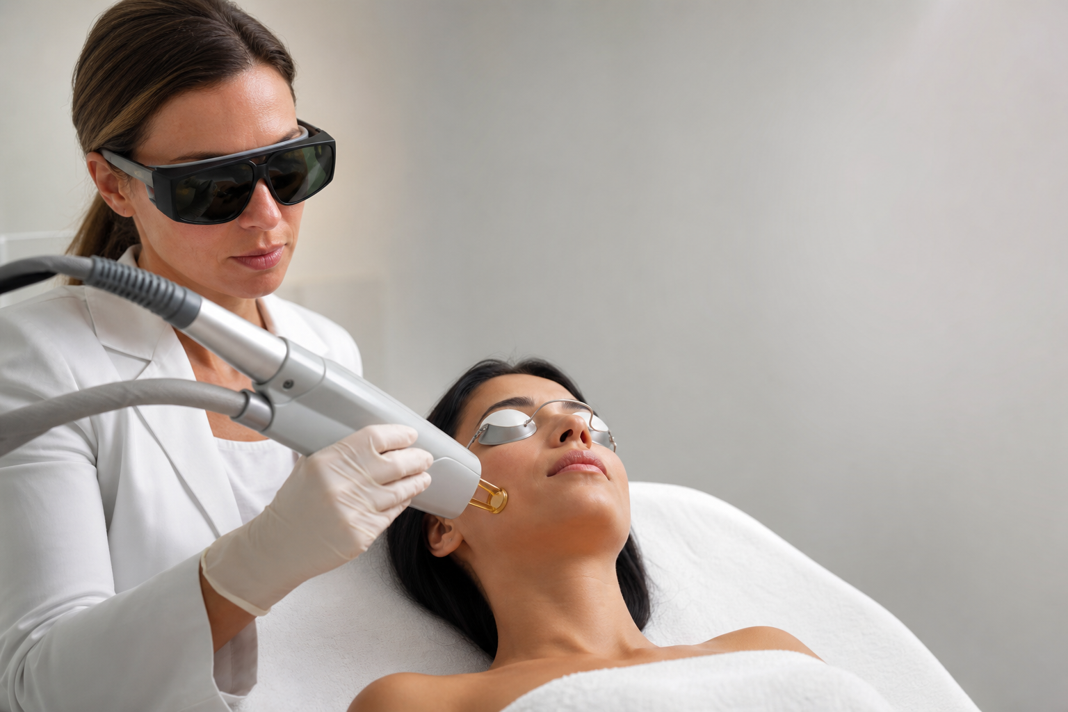
A typical Alexandrite laser session begins with skin cleansing and application of topical anesthesia if indicated for comfort. Protective eyewear is mandatory for both patient and practitioner. The practitioner then delivers controlled laser pulses over the pigmented lesions, often perceived as brief snapping sensations akin to a rubber band flick.
Post-procedure, patients might notice transient redness and swelling akin to mild sunburn. Pigmented spots may darken initially before gradually lightening and fading over days to weeks. Multiple treatment sessions are often necessary for optimal clearance, spaced to allow skin recovery and pigment resolution.
Continuous sun protection and avoidance of exacerbating factors post-treatment are critical to sustaining results. Patients should have realistic expectations, understanding that complete pigment removal may not always be achievable, particularly for lesions with complex or deep pigmentation.
How Does the Alexandrite Laser Compare with Other Lasers for Pigmentation Treatment?
The landscape of laser treatment for pigmented lesions includes a variety of devices, each with unique wavelengths and indications. Compared to Q-Switched Nd:YAG lasers and Picosecond lasers, Alexandrite lasers offer a highly melanin-selective 755 nm wavelength optimizing them for epidermal pigment removal on lighter skin types.
Q-Switched Nd:YAG lasers operate at 1064 nm, penetrating deeper and being safer for darker skin due to reduced melanin absorption. Picosecond lasers deliver ultrashort pulses enabling enhanced pigment fragmentation with possibly fewer treatments. Ruby lasers at 694 nm also target melanin but have largely been supplanted by Alexandrite lasers due to adjustable pulse durations and better skin compatibility.
Fractional and Er:YAG lasers, while valuable for resurfacing, photodamage, and improving skin texture, are adjuncts rather than primary pigment-specific treatments. Selecting the appropriate laser modality requires comprehensive lesion evaluation and patient skin type consideration, often guided by references such as "Which Lasers Are Used to Treat Pigmentation?" and "Laser Treatments for Pigmentation: A Complete Guide to Types of Dark Spots, Lasers, Results and Risks."