Understanding why melanin reacts to laser energy is fundamental in the nuanced field of dermatological and aesthetic laser treatments. This concept underpins procedures such as laser tattoo removal, hair removal, skin rejuvenation, and pigment correction. Melanin, the natural pigment that confers color to our skin, hair, and eyes, plays a pivotal role in how our skin interacts with laser light. These interactions dictate not only treatment outcomes but also the safety and risk profiles, especially for individuals with darker or tanned skin.
This article is designed for a broad audience—clients considering laser treatments, aesthetic professionals, and dermatologists alike. It aims to demystify the science behind melanin's response to laser energy, clarify expected skin reactions, illuminate associated risks, and furnish practical guidance on prevention and early assessment of adverse effects. With particular emphasis on the variability introduced by skin type and pigmentation levels, this discussion is vital for informed decision-making in laser procedures.
Laser treatments have become increasingly popular due to their precision and efficacy. However, melanin’s variable absorption of laser wavelengths introduces complexities that can influence both effectiveness and safety. This complexity necessitates a detailed understanding of how laser settings and skin characteristics interplay to either maximize therapeutic benefits or inadvertently cause skin damage. This article explores these dynamics extensively to support optimal laser treatment strategies.
What Is Melanin and How Does It Function in the Skin?
Melanin is a bio-pigment responsible for the diverse range of human skin tones, hair colors, and eye pigmentation. Its primary biological role is protective: by absorbing ultraviolet (UV) radiation, melanin helps shield underlying skin cells from DNA damage that can lead to conditions including premature aging and skin cancer.
Two major types of melanin exist: eumelanin and pheomelanin. Eumelanin imparts brown to black hues and is more effective at photoprotection, whereas pheomelanin is associated with red and yellow tones and offers comparatively less defense against UV rays. The relative abundance and distribution of these melanin types vary among individuals and contribute to differing Fitzpatrick skin phototypes.
Melanin resides predominantly in the basal layer of the epidermis within specialized cells called melanocytes, and is distributed into surrounding keratinocytes. Its light-absorbing properties, while protective against the sun’s harmful rays, also render it a chromophore that can absorb energy from laser light. This dual role is central to understanding melanin’s interaction with laser energy during skin treatments.
Why Does Melanin React to Laser Energy? The Fundamental Physical Principles
The reaction of melanin to laser energy is governed by its intrinsic optical absorption characteristics. Lasers emit highly specific wavelengths of coherent light that target chromophores—molecules within the skin that absorb light energy—to induce therapeutic effects. Melanin is a prime chromophore due to its broad absorption spectrum extending from ultraviolet through visible to near-infrared wavelengths.
When exposed to laser light at an appropriate wavelength, melanin absorbs the energy and converts it into heat through a process called photothermolysis. The generated heat can selectively damage the melanocytes or melanin-containing structures targeted in a treatment. For example, in tattoo removal, laser energy breaks down tattoo pigment particles so the body can phagocytose and eliminate them.
The key to effective laser therapy is balancing this absorption: sufficient to achieve the desired effect on pigmented targets while minimizing collateral damage to surrounding healthy tissue. If laser settings or skin conditions lead to excessive melanin absorption, undesirable outcomes such as burns, pigmentary changes, or inflammation may occur, especially in skin types with higher baseline melanin content.
Which Skin Types Are Most Vulnerable to Laser-Induced Melanin Reactions?
Skin vulnerability during laser treatments strongly correlates with the Fitzpatrick skin phototype, which classifies skin from I (very fair) to VI (very dark) based on pigmentation and response to UV exposure. Individuals with lighter skin (phototypes I to III) have lower melanin density and generally tolerate laser treatments with a reduced risk of pigment alteration, though they remain susceptible to other side effects such as burns or scarring if improperly treated.
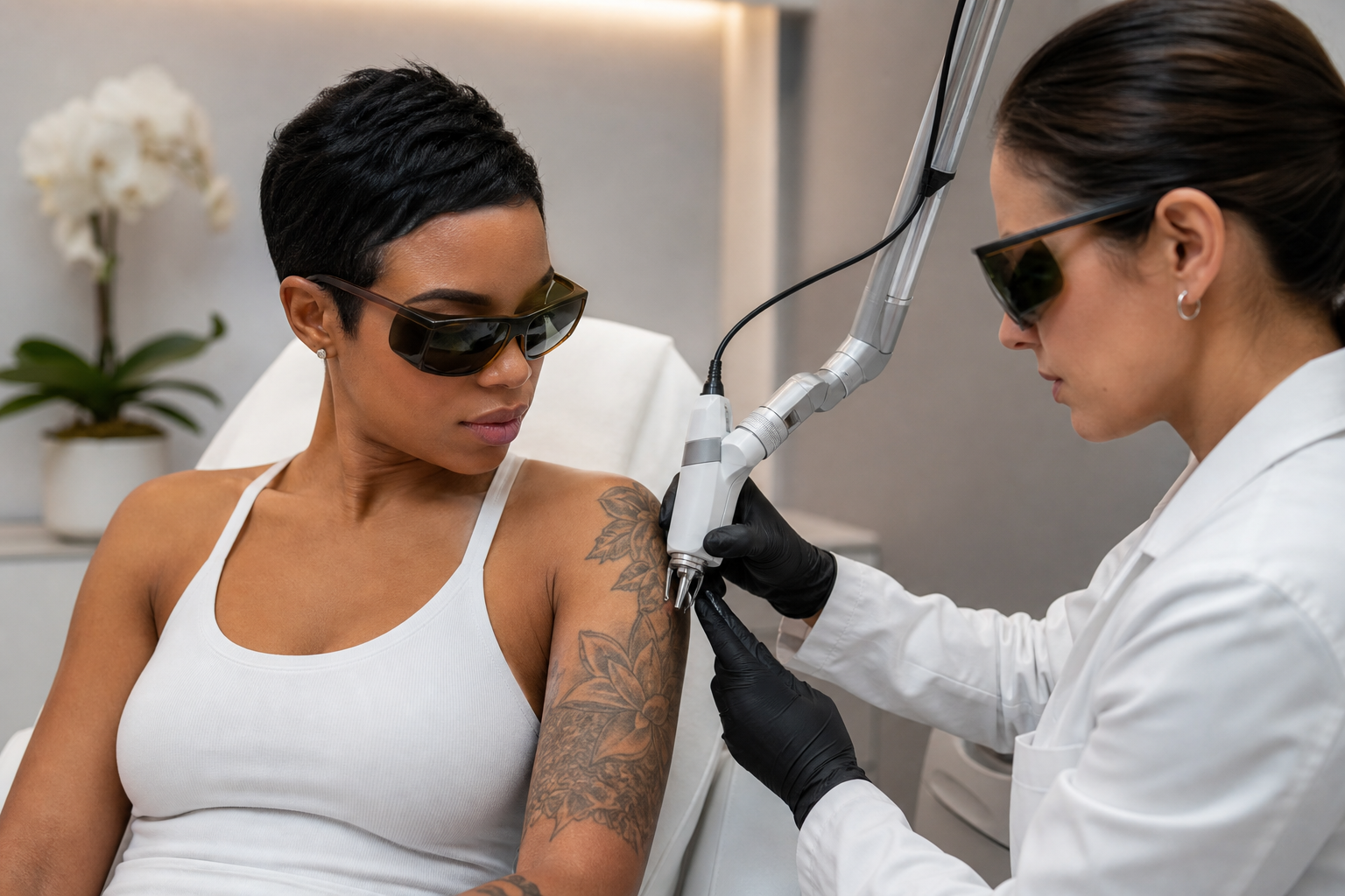
Conversely, darker skin types (phototypes IV to VI) harbor significantly more melanin within the epidermis and dermis. This elevated melanin concentration amplifies laser energy absorption, raising the risk of adverse effects such as post-inflammatory hyperpigmentation, hypopigmentation, thermal injury, or scarring if laser parameters are not judiciously selected.
Additionally, recent tanning increases epidermal melanin, transiently intensifying these risks. Hence, individuals with tanned skin require postponed treatments until the skin tone normalizes. This stratification is crucial when planning laser tattoo removal or pigmentation treatments to ensure safety and efficacy.
How Does Melanin Specifically React During Laser Tattoo Removal?
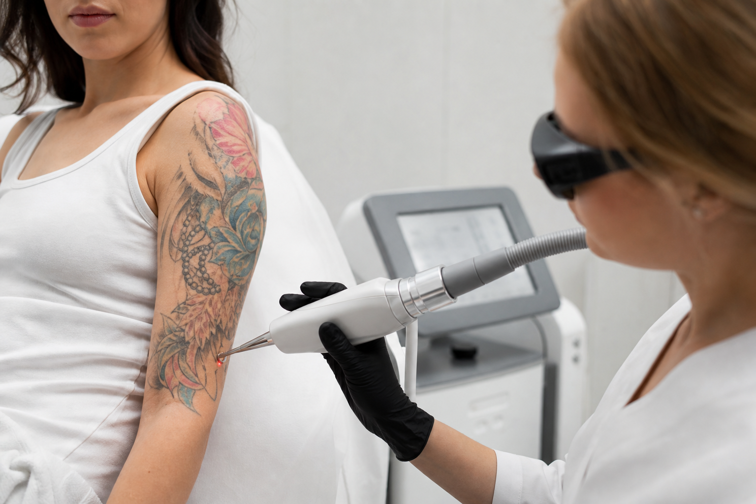
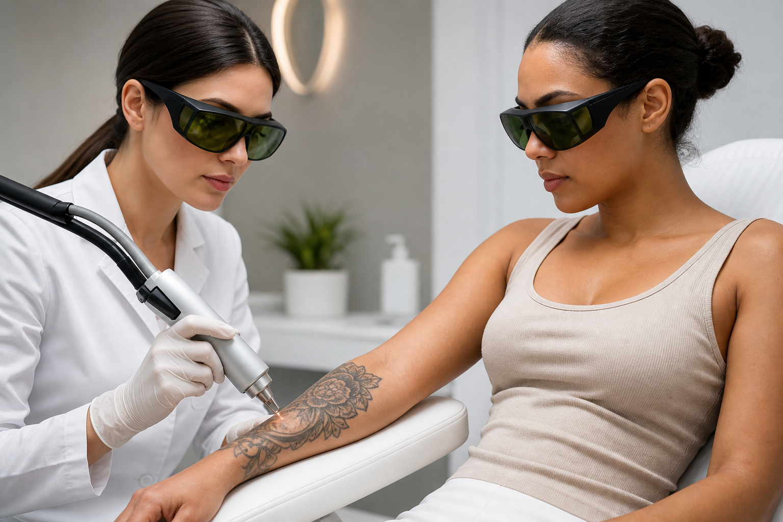
Laser tattoo removal relies on selective photothermolysis targeting the ink particles embedded in the dermis. Tattoo pigments, often darker and more optically dense than surrounding melanin, absorb laser energy more readily. However, epidermal melanin also absorbs some energy, which necessitates precise laser wavelength and fluence selection to preferentially affect the tattoo ink while preserving healthy skin.
In treatments involving darker skin or recently tanned individuals, there is a heightened risk that melanin in the epidermis absorbs excessive laser energy, leading to inflammation, burns, and pigmentary dysregulation. Such complications underscore the importance of using lasers with longer wavelengths, like the Nd:YAG 1064 nm, that preferentially penetrate deeper tissues while exerting less energy absorption by epidermal melanin.
Moreover, laser parameters such as pulse duration and fluence must be meticulously calibrated. Short pulses limit heat diffusion, reducing damage to non-target tissues. Treatment intervals allowing skin recovery also minimize cumulative injury and pigment alterations.
What Are the Common Risks Associated with Melanin-Laser Interactions and How Can They Be Recognized?
Risks stemming from melanin's absorption of laser energy manifest in several ways. Hyperpigmentation, characterized by darkening of the skin at or near the treatment site, is a common response due to increased melanocyte activity or melanin retention following inflammatory injury.
Conversely, hypopigmentation involves loss of pigment in treated skin areas, often resulting from melanocyte damage or destruction. Thermal injuries such as burns and blistering may also arise if laser energy exceeds the skin’s tolerance, especially in heavily pigmented skin where melanin absorbs high energy levels.
Normal post-treatment erythema and edema should subside within days; however, persistence or worsening of redness, pain, blistering, or pigmentary changes beyond the typical healing timeframe warrants clinical evaluation. Early recognition of these signs facilitates timely intervention to mitigate long-term sequelae.
How Can These Risks Be Minimized During Laser Procedures?
Preventing adverse outcomes in laser treatments involving melanin requires a comprehensive approach beginning with accurate skin type assessment using the Fitzpatrick scale and evaluation of recent sun exposure or tanning status. This information guides laser selection and parameter settings tailored to individual skin characteristics.
Utilizing lasers emitting longer wavelengths—such as the Nd:YAG laser at 1064 nm—can reduce epidermal melanin absorption, thereby lowering the risk of pigmentary complications in darker skin types. Employing appropriate fluence and pulse durations further refines energy delivery to target chromophores while sparing surrounding tissue.
Additionally, practitioners should advise patients to avoid sun exposure, tanning beds, and self-tanning products for weeks before and after treatment to stabilize melanin concentration and enhance treatment safety. Post-treatment protocols emphasizing skin hydration, inflammation control, and rigorous photoprotection are essential components of risk management.
Why Do Experts Recommend Postponing Laser Treatments on Tanned Skin?
Tanned skin exhibits a temporary surge in epidermal melanin, amplifying laser energy absorption and elevating the risk of burns, pigmentary changes, and prolonged inflammation. This heightened melanin presence can unpredictably alter laser-tissue interactions, compromising treatment efficacy and safety.
Consequently, laser treatments, particularly tattoo removal and pigment-targeting procedures, are generally deferred until the patient’s skin returns to its baseline pigmentation. The duration of this waiting period varies depending on the intensity of pigmentation and the individual’s skin biology, often ranging from several weeks to a few months.
This conservative approach reduces complication rates and enhances overall treatment outcomes, underscoring a critical safety principle in laser dermatology highlighted in guidance documents such as Why Is Tattoo Removal Postponed on Tanned Skin?
When Should Patients Seek Professional Evaluation Following a Laser Treatment?
Patients should remain vigilant for signs indicative of possible complications and seek professional assessment if they experience prolonged or worsening redness, blister formation, intense pain, unusual pigmentation changes extending beyond treated areas, or symptoms suggestive of infection such as pus, fever, or increased swelling.
Early consultation enables clinicians to institute suitable interventions—ranging from topical corticosteroids to antimicrobial therapy—to prevent permanent pigmentary alterations or scarring. Close follow-up is especially recommended for individuals with darker skin, whose higher melanin content predisposes them to more pronounced reactions.
How Does Fitzpatrick Skin Typing Guide Safe and Effective Melanin-Targeted Laser Treatments?
The Fitzpatrick skin type classification serves as a cornerstone in tailoring laser therapies. By categorizing skin based on inherent pigmentation and UV reactivity, it informs the selection of laser wavelength, pulse duration, and energy settings to optimize treatment precision while mitigating risks.
Light skin types (I–III) generally tolerate a broader spectrum of laser parameters, although careful adjustment remains necessary to avoid burns or pigmentary alterations. Darker skin types (IV–VI) necessitate cautious settings favoring longer wavelengths and lower fluences, exploiting deeper laser penetration and minimizing absorption by epidermal melanin.
Understanding this classification enables practitioners to predict potential responses and adapt protocols accordingly, promoting patient safety and satisfaction. Further insights can be found in resources such as What Is the Fitzpatrick Skin Type and Why Does It Matter?
Why Is It Essential to Comprehend Melanin’s Reaction to Laser Energy?
Comprehending melanin’s interaction with laser energy is indispensable for optimizing clinical outcomes and safeguarding patient well-being. This knowledge facilitates personalized treatment planning that harmonizes efficacy with safety, underscores the rationale behind using specific laser types and settings, and guides patient selection and education.
As non-invasive aesthetic and medical laser procedures proliferate, the demand for meticulous protocols addressing melanin-related risks grows concomitantly. Enhancing awareness among patients and professionals fosters realistic expectations, diligent adherence to preventive measures, and prompt management of complications—ultimately refining therapeutic experiences.