Dark spots on the skin, often referred to as hyperpigmentation, are a common cosmetic concern that many individuals confront at some point in their lives. These spots, which range from small freckles to larger patches, can appear on the face, hands, or other areas frequently exposed to sunlight. For many, dark spots primarily represent a cosmetic nuisance, affecting self-esteem and the overall perception of one's appearance. However, it is critical to recognize that not all pigmentation changes are purely aesthetic; some may signify underlying medical conditions that necessitate professional evaluation.
This article explores the crucial question: When is a dark spot more than a cosmetic concern? Specifically, it addresses individuals noticing pigmentation changes who seek to understand when these marks are harmless and when they could be indicative of more serious health issues. Understanding this distinction is vital not only for appropriate treatment decisions but also for safeguarding overall skin health. The topic also has significant relevance for those considering laser treatments as a solution for pigmentation, emphasizing the importance of thorough dermatologic assessments before such interventions.
By delving into the nature of dark spots, their causes, and when to seek medical advice, this editorial aims to offer clarity and guidance for patients and practitioners alike. The following sections unpack the factors that distinguish benign pigmentation from pathological changes, detail the diagnostic approaches necessary before laser therapy, and highlight common concerns and best practices connected to treating dark spots.
What Are Dark Spots and Why Do They Occur?
Dark spots, or hyperpigmentation, occur when melanin—the pigment responsible for skin color—is produced in excess within certain areas. This phenomenon results in noticeable patches or flecks that appear darker than the surrounding skin. Several factors contribute to hyperpigmentation, including prolonged sun exposure, hormonal fluctuations, skin inflammation, injury, medication side effects, and various systemic health conditions.
The most commonly encountered types of dark spots include solar lentigines (often called sun spots or age spots), melasma, and post-inflammatory hyperpigmentation (PIH). Solar lentigines typically arise from chronic ultraviolet (UV) radiation damage, presenting as harmless yet persistent brown spots usually in sun-exposed areas. Melasma, predominantly affecting women, is driven by hormonal influences and often manifests as symmetric, blotchy patches on the cheeks, forehead, and upper lip. On the other hand, PIH results from skin trauma or inflammation, such as acne or eczema, leading to localized pigment darkening following the resolution of the primary condition.
Understanding these distinct types of pigmentation is essential in selecting adequate and safe treatment options. While many dark spots are benign and primarily a cosmetic issue, some pigmentation changes represent deeper or more serious alterations requiring professional evaluation.
When Could a Dark Spot Indicate a More Serious Skin Condition?
While the majority of dark spots carry no serious health consequences, vigilance is necessary as some may indicate premalignant or malignant skin conditions. One critical example is melanoma, the most dangerous form of skin cancer, which can initially mimic benign pigmented lesions. Changes such as rapid growth, color variation, irregular borders, asymmetry, bleeding, or itchiness in an existing spot—or the sudden appearance of a new lesion—should prompt immediate dermatologic assessment.
Beyond malignancies, certain pigment alterations might be secondary to internal disorders. Hormonal imbalances, as seen in conditions like polycystic ovary syndrome or adrenal gland disorders, can trigger abnormal pigmentation patterns. Chronic inflammatory diseases may also cause persistent hyperpigmentation signaling ongoing systemic issues. Recognizing when a dark spot reflects these underlying conditions is paramount to effective management and overall health preservation.
Therefore, while many pigmentation issues are manageable cosmetic concerns, specific warning signs mandate prompt professional attention to exclude or diagnose serious skin or systemic pathology.
How Can One Differentiate Between Harmless and Concerning Pigmentation?
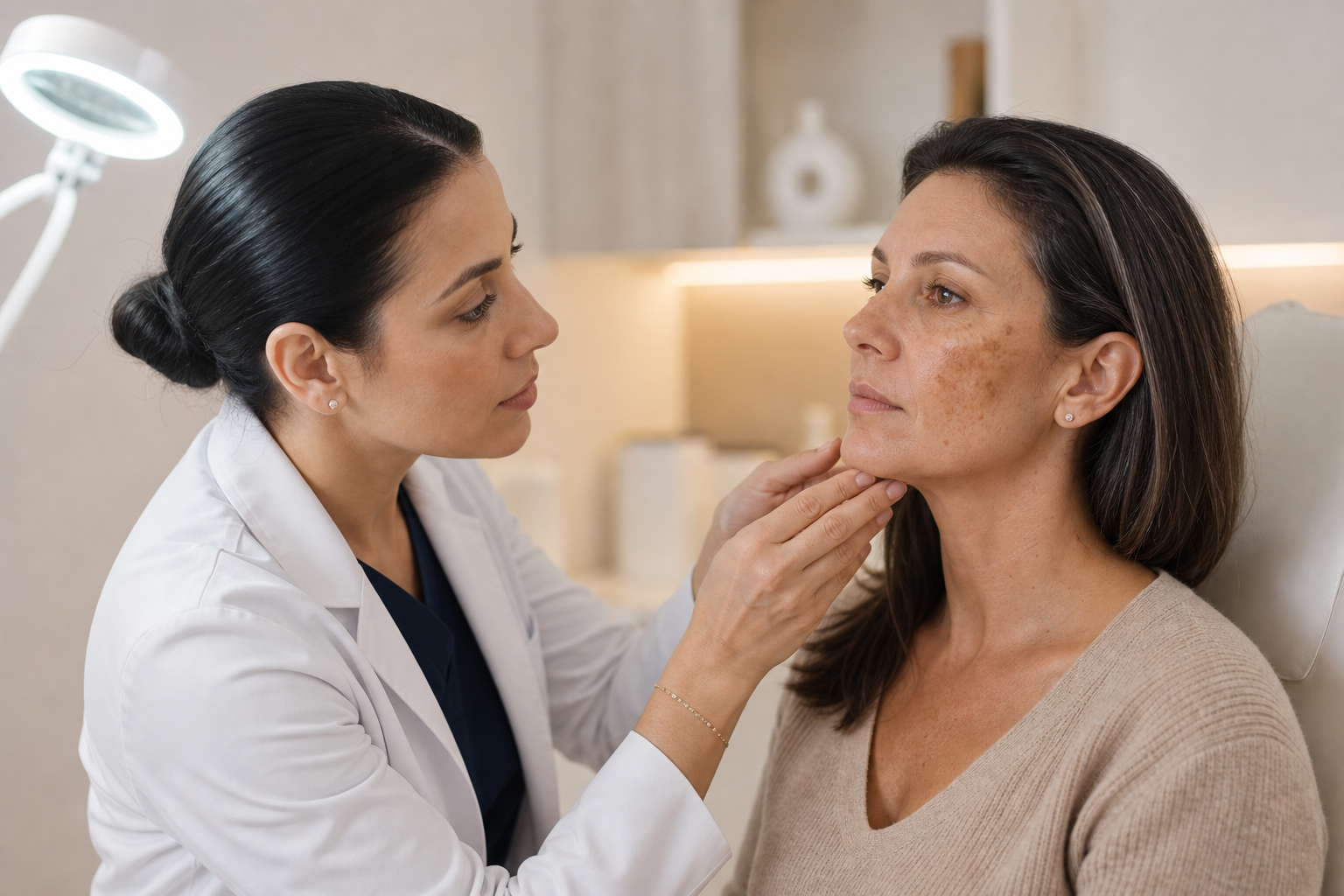
Self-assessment of dark spots is inherently limited and can lead to misdiagnosis or inappropriate treatment. Dermatologic evaluation remains the gold standard in distinguishing benign lesions from those requiring intervention. At the initial consultation, a dermatologist performs a detailed visual examination, often employing enhanced diagnostic tools such as Wood's lamp — a specialized ultraviolet light that reveals nuances in pigmentation depth and pattern not visible under normal lighting.
This approach allows clinicians to differentiate epidermal hyperpigmentation, which is superficial and typically more responsive to laser or topical treatments, from dermal pigmentation residing deeper in the skin—often more resistant to standard therapies. Additionally, expert evaluation helps clarify whether pigmentation is a sequela of inflammation, hormonal influence, or a suspicious lesion indicative of malignancy.
Such comprehensive assessment mitigates risks associated with inappropriate interventions, such as aggravation of the lesion or concealment of serious conditions.
Why Is Dermatologic Assessment Essential Before Laser Treatment?
Laser therapy has gained significant popularity as an effective means of addressing various pigmentation disorders, offering benefits like targeted melanin breakdown and skin tone harmonization. Nonetheless, this modality requires prudent patient selection and meticulous pre-treatment evaluation to ensure safety and efficacy.
Applying laser treatment without a definitive diagnosis risks adverse outcomes, including exacerbation of pigmentation, skin irritation, or even stimulating latent malignant processes. A dermatologist’s assessment informs whether a lesion is suitable for laser intervention and guides the choice of laser type and parameters based on pigment depth and skin type.
For example, epidermal pigmentation responds well to Q-switched or Alexandrite lasers, while dermal pigmentation may necessitate fractional laser approaches or alternative therapies. Furthermore, certain lesions, like melanocytic nevi or actinic keratoses, are contraindications for laser removal due to malignancy risk. In these contexts, the dermatologist may recommend biopsy or other measures instead.
What Are the Risks of Immediate Laser Treatment Without Prior Evaluation?
Engaging in laser treatment without a proper clinical diagnosis can trigger unintended complications. Pigmentation may paradoxically worsen, especially in individuals prone to post-inflammatory hyperpigmentation. Incorrect laser parameters can cause burns, scarring, or uneven skin tone. Moreover, treating lesions of uncertain benignity without histologic confirmation may delay diagnosis of potentially fatal diseases like melanoma.
There is also the concern that hormonal or inflammation-driven pigmentation might persist or recur if the underlying cause is unaddressed, rendering laser treatment less effective or even counterproductive. Consequently, deferring to specialist evaluation protects against these pitfalls and optimizes therapeutic outcomes.
How Do Hormonal Changes Influence Dark Spots, and What Implications Do They Have for Treatment?
Hormonal pigmentation, particularly melasma, presents distinct challenges. Triggered by estrogen and progesterone levels during pregnancy, oral contraceptive use, or hormone replacement therapy, melasma involves activation of melanocytes resulting in diffuse, symmetrical patches commonly on the face.
This condition often fluctuates with hormonal status and may improve post-pregnancy or after adjusting medications. Therefore, aggressive laser treatments during active hormonal influence risk exacerbation. Dermatologists frequently advocate for integrated management, including hormonal evaluation, topical agents like hydroquinone or tranexamic acid, sun protection, and cautious laser use timed to hormonal stability.
Recognizing hormonal triggers and counseling patients accordingly prevents unnecessary frustration and side effects from premature or solitary laser treatments.
What Can Patients Typically Expect During and After Laser Treatment for Dark Spots?
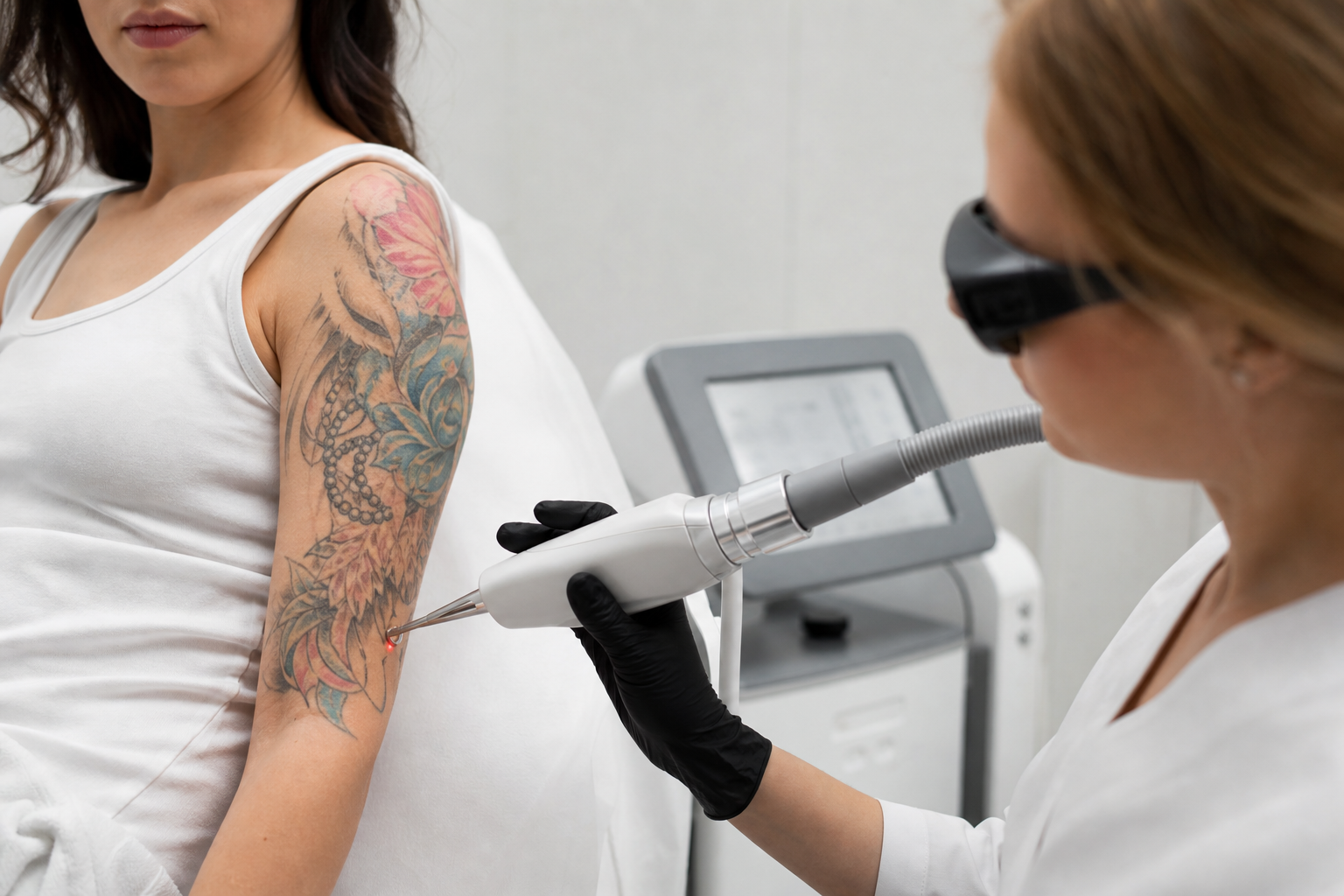
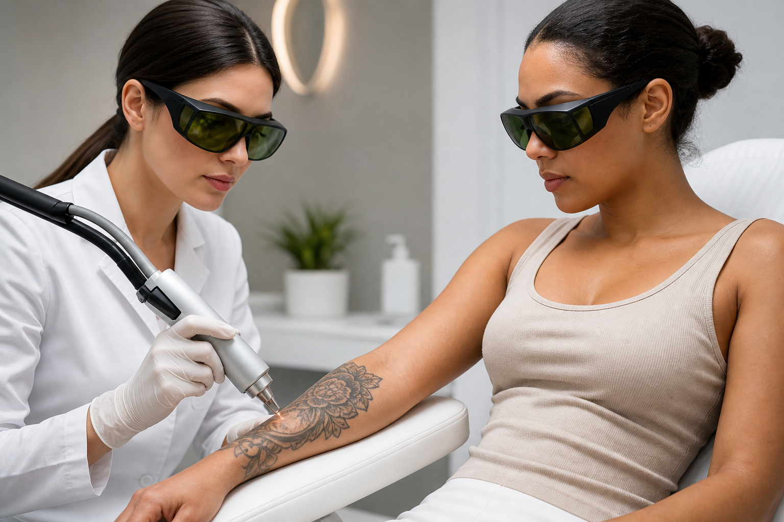
Following dermatologic diagnosis, laser treatment for pigmentation is often planned over multiple sessions to achieve gradual improvement. Choices among Q-switched lasers, Alexandrite, or fractional devices depend on pigmentation type and depth. Each session targets melanin granules to encourage gradual dispersal and clearance.
Post-treatment skin may exhibit redness, mild swelling, and peeling as part of the natural healing process. These effects generally resolve within days. Strict sun avoidance and application of broad-spectrum sunscreen are critical to prevent rebound pigmentation. Patients typically observe differences after initial treatments; however, sustained benefits rely on ongoing skin care, lifestyle modifications, and sometimes maintenance therapies.
Open communication with the dermatologist throughout this process ensures expectations are aligned and any adverse effects are promptly addressed.
Common Questions Addressed About Dark Spots and Their Management
Does every dark spot require treatment? No, many pigmented lesions are benign and pose no health threat. Treatment is optional unless the spot causes distress or cosmetic dissatisfaction.
How do I know if a pigmented spot is dangerous? Warning signs include rapid growth, asymmetry, color changes, irregular borders, bleeding, or persistent itching. Such symptoms warrant urgent professional evaluation.
Is laser treatment safe? When performed by a qualified dermatologist following comprehensive diagnosis, laser treatments are generally safe. Incorrect use or unsupervised procedures increase risks.
Can dark spots be permanently removed? Most benign pigmentation can be significantly reduced or removed, though complete permanence varies based on individual skin factors and ongoing exposures.