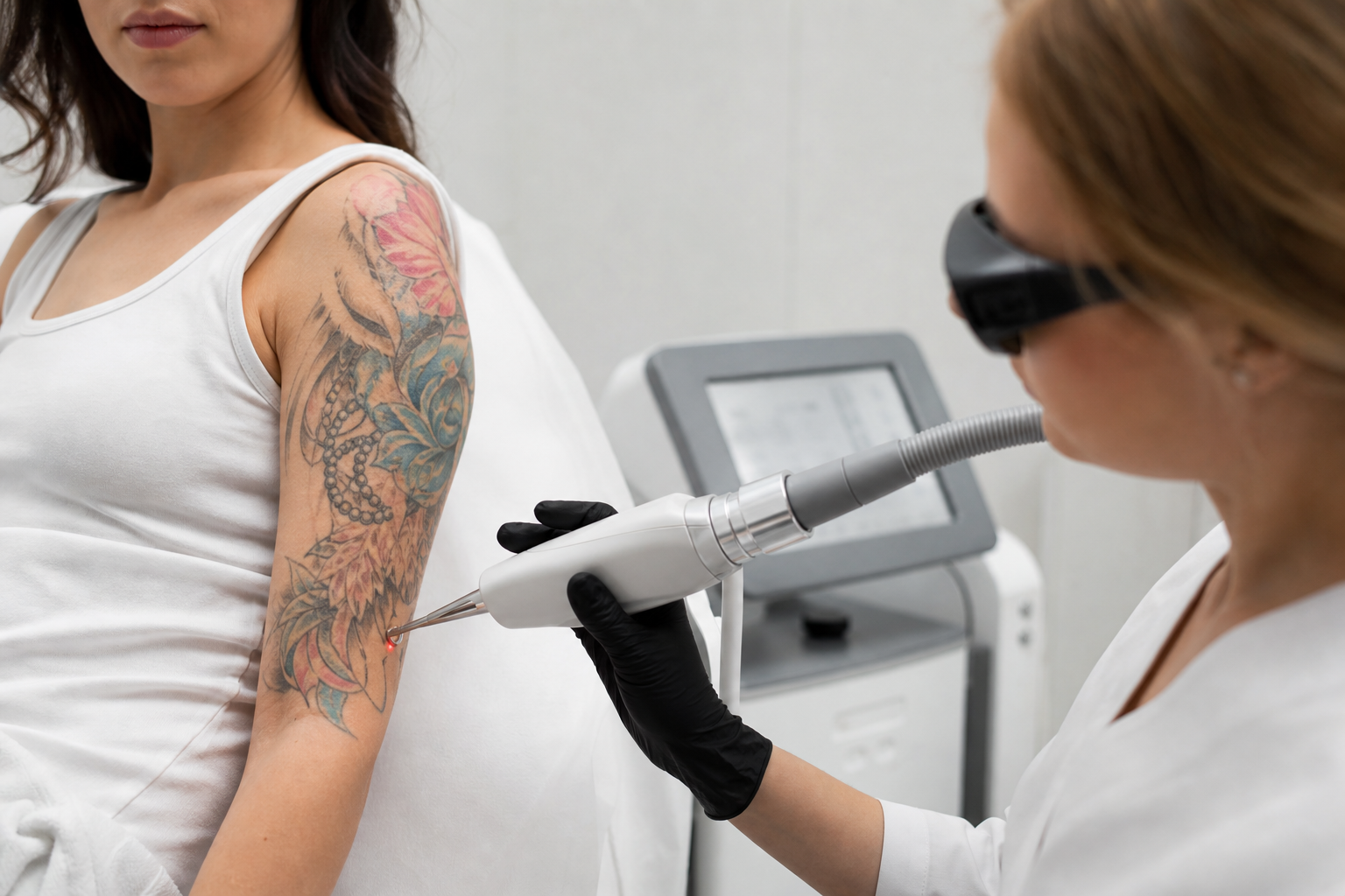
Understanding pigmentation changes in the skin is crucial for effective and safe treatment, particularly when considering laser therapies. Pigmentation refers to areas of skin where melanin—the natural pigment responsible for color—is concentrated, resulting in darker spots or patches. However, not all pigmentations are alike; they differ fundamentally in where the pigment lies within the skin layers. The primary distinction exists between epidermal pigmentation, which occurs in the skin’s outermost layer, and dermal pigmentation, which is found deeper beneath the surface. Recognizing these differences is essential for dermatologists, skincare professionals, and patients alike, as it directly influences treatment choices, anticipated outcomes, and the safety of interventions such as laser therapy.
This article offers a detailed exploration of epidermal versus dermal pigmentation, emphasizing why the depth of pigment profoundly changes treatment strategies. We will clarify distinguishing features of these pigmentations without encouraging self-diagnosis and highlight when a thorough dermatologic assessment is indispensable before any laser procedures. For individuals seeking clarity on their pigmentation concerns or professionals refining their approach to pigment correction, this in-depth guide elucidates the complexities and helps navigate the path to improved skin health.
In an age dominated by quick cosmetic fixes and abundant online information, grasping the nuances of pigment depth transcends mere aesthetic interest. It is fundamentally about the well-being and longevity of one's skin. Join us as we unpack the clinical, diagnostic, and therapeutic aspects of pigmentation, with particular attention to laser treatments and their relationship with pigment depth.
What Distinguishes Epidermal Pigmentation and How Can It Be Identified?
Epidermal pigmentation occurs when melanin accumulates in the epidermis, the uppermost layer of the skin. This kind of pigmentation typically manifests as dark spots with clear boundaries and is often attributable to sun exposure—such as solar lentigines or freckles—or hormonal influences, as seen in melasma. These pigmentations predominantly appear on sun-exposed areas like the face.
Identifying epidermal pigmentation involves looking at the lesion’s color and response to diagnostic tools like the Wood’s lamp, which emits ultraviolet light. Pigmentations situated superficially in the epidermis usually fluoresce brightly under this lamp, helping to differentiate them from deeper pigmentations. Because the pigment lies near the skin surface, epidermal pigmentation generally responds well to laser treatments. The lighter and more superficial nature of these spots often allows for effective laser penetration and quicker recovery times.
The skin’s natural renewal cycle plays a significant role in addressing epidermal pigmentation. Given its location in the epidermis, the skin cells responsible for pigment production and accumulation undergo regular turn-over, which supports recovery and restoration of even skin tone post-treatment. This regenerative capacity makes epidermal pigmentation less invasive to treat and typically associated with fewer complications.
What Are the Characteristics of Dermal Pigmentation and Its Clinical Presentation?
In contrast, dermal pigmentation involves pigment deposits located within the dermis, the thicker, connective tissue-rich layer beneath the epidermis. The deeper location produces lesions that may appear blue, gray, or yellowish, often with fuzzy or indistinct edges. Examples include certain scar-related pigmentations, post-inflammatory hyperpigmentation penetrating below the epidermis, and some variants of melasma with a significant dermal component.
Clinically, dermal pigmentations can be more challenging both to diagnose and to treat. Unlike epidermal pigmentations, they do not fluoresce clearly under Wood’s lamp because ultraviolet light does not reach the deeper dermis effectively. This limitation often leads to diagnostic uncertainty and complicates the selection of therapeutic modalities.
Successfully managing dermal pigmentation often necessitates a comprehensive examination by a dermatologist, potentially involving skin biopsies or advanced imaging techniques to ascertain the exact depth and nature of the pigmentation. Treatments are generally more complex, frequently requiring a combination of approaches such as deep-penetrating lasers (e.g., Q-switched or fractional lasers), chemical peels, or even intradermal injections. Preventive strategies, including stringent sun protection and post-procedural skincare, also play pivotal roles in maintaining treatment benefits and minimizing recurrence.
Why Does Pigment Depth Dictate the Choice of Treatment?
The depth at which pigment resides within the skin is a fundamental determinant when planning pigment correction therapies, particularly laser interventions. Lasers target melanin pigment by delivering light energy absorbed and converted to heat, thereby disrupting pigment granules. However, laser efficacy is intrinsically linked to how deeply the pigment sits beneath the skin surface.
Epidermal pigmentations, being superficial, allow laser energy to focus precisely on the pigment with minimal collateral damage. This accessibility results in effective breakdown of pigment particles and subsequent elimination through the skin’s natural processes. Conversely, dermal pigmentations require lasers with wavelengths and penetrative capacities sufficient to reach the deeper dermis. Such treatments inherently carry increased risk, as deeper laser energy can harm surrounding tissues, potentially causing inflammation, scarring, or unwanted pigmentation changes.
Furthermore, deep pigmentations often demand a staged or multifaceted treatment approach. Immediate and aggressive therapy is rarely advisable; instead, dermatologists may combine several modalities over time to achieve satisfactory results while minimizing adverse effects. Therefore, an accurate understanding of pigment depth is essential to balance treatment efficacy and patient safety.
When and Why Should You Seek Dermatological Evaluation Before Laser Treatment for Pigmentation?
Dermatological assessment is vital in multiple scenarios prior to laser treatment of pigmentation. Foremost, a professional diagnosis verifies the pigment type and its precise depth within the skin layers. This evaluation ensures that the chosen treatment aligns with the nature of the pigmentation and avoids potentially harmful interventions.
Additionally, pigmentation changes that evolve in size, shape, or color warrant urgent medical evaluation to rule out malignant or precancerous conditions, which are contraindications for cosmetic laser treatments. An expert dermatologist employs tools such as dermatoscopy, Wood’s lamp examination, and when necessary, skin biopsies to differentiate benign pigmentations from dermatological disorders or skin cancers.
Without professional examination, patients risk undertaking inappropriate treatments that may exacerbate their condition or mask underlying disease. Therefore, consulting a dermatologist safeguards long-term skin health and optimizes aesthetic outcomes. Experts may also recommend alternative or adjunctive therapies, particularly in cases of hormonal pigmentations or chronic conditions where laser therapy alone may be insufficient or inadvisable.
How Do Different Laser Treatments Interact with Pigmentation Depth?
Laser technology relies on variable wavelengths and pulse durations to target melanin selectively. Shorter-wavelength lasers with limited penetration are typically reserved for treating epidermal pigmentations. These lasers concentrate energy close to the surface, allowing precise targeting of superficial pigment without damaging deeper structures.
For dermal pigmentation, lasers capable of deeper skin penetration are employed, such as Q-switched Nd:YAG lasers or fractional lasers. These devices deliver energy that reaches the dermis, breaking up pigment particles embedded therein. Nevertheless, due to the increased risk of damaging surrounding tissue, treatments must be carefully calibrated and often entail prolonged treatment courses.
Patient variables—including skin phototype, age of pigmentation, and previous treatments—further influence laser selection. Improper laser use, especially on deeper pigmentations or darker skin tones, can result in post-inflammatory hyperpigmentation or hypopigmentation, potentially worsening the cosmetic appearance. Hence, personalization of laser parameters under specialist supervision is paramount.
What Non-Laser Alternatives Exist for Treating Deep Dermal Pigmentation?
When laser therapy is unsuitable or limited, especially for deep dermal pigmentations, alternative treatments gain importance. Chemical peels that penetrate to medium or deep skin layers can exfoliate pigmented cells and stimulate regeneration, effectively reducing pigmentation.
Microneedling is another adjunct technique that promotes collagen production and enhances the skin’s ability to absorb topical agents. Coupled with skin-lightening products containing agents like hydroquinone, azelaic acid, or retinoids, these methods can gradually lighten persistent pigmentations. However, such treatments require patient adherence, professional monitoring, and diligent sun protection to prevent relapse.
Ultimately, combination therapies under dermatological guidance often yield superior results by targeting pigment at multiple levels through complementary mechanisms.
How Can You Determine If a Pigmentation Spot Requires Medical Attention Rather Than Cosmetic Procedures?
Not all pigmented skin lesions are purely cosmetic. Signs that warrant immediate medical evaluation include rapid changes in the lesion’s size or color, irregular borders, varied pigmentation within one spot, ulceration, itching, or bleeding. These features may indicate melanoma or other serious dermatological conditions that demand prompt diagnosis and treatment.
Moreover, pigmentation emerging after skin injury, drug use, or hormonal fluctuations should be medically examined to assess the underlying causes. Dermatologists can differentiate benign pigmentary changes from conditions that necessitate medical intervention, ensuring appropriate and safe management.
Regular skin checks, particularly for individuals with extensive sun exposure, a history of skin cancer, or notable pigmentary changes, are essential preventative measures to detect malignant transformations early.