In the world of dermatology and aesthetic treatments, precise assessment of skin conditions is crucial for effective and safe interventions. One diagnostic tool that has gained significant importance in evaluating pigmentation disorders is the Wood's lamp. This specialized lamp employs a specific wavelength of ultraviolet (UV) light to illuminate the skin, revealing patterns and depths of pigmentation that are often imperceptible to the naked eye. Understanding what a Wood's lamp is, how it functions, and how it assists in assessing pigmentation is essential for patients considering pigmentation treatments and clinicians striving for optimal therapeutic outcomes.
Wood's lamp examination serves as a non-invasive, rapid, and insightful diagnostic method particularly valuable for individuals experiencing issues such as hyperpigmentation, melasma, solar lentigines (sun spots), and other pigmentary irregularities. It also holds a pivotal role in the preparatory assessment before laser treatments aimed at correcting pigmentation concerns. This examination not only aids in diagnosing but also in differentiating between various pigmentary conditions—knowledge that is vital to avoid ineffective or potentially harmful treatment choices.
In this comprehensive editorial, we will explore the fundamentals of Wood's lamp technology, delve into its application in pigmentation analysis, and clarify its indispensable role within dermatologic evaluation before laser interventions. By the end, readers will appreciate how this technology bridges the gap between clinical examination and tailored treatment strategies for pigmentation disorders.
What Is a Wood's Lamp and How Does It Function in Dermatology?
A Wood's lamp is a diagnostic device named after Robert Williams Wood, an American physicist who first described its use in the early 20th century. It emits ultraviolet light at a wavelength around 365 nanometers, which is within the UVA spectrum. This specific wavelength is significant because it causes certain substances and changes in the skin to fluoresce or emit visible light, thereby exposing characteristics invisible under standard lighting conditions.
The principle behind the Wood's lamp examination lies in the interaction of UV light with components of the skin such as melanin, collagen, bacteria, and various epidermal alterations. When the lamp is directed at the skin in a darkened environment, different pigmentation types and skin conditions emit distinct fluorescence colors. For example, areas of superficial epidermal hyperpigmentation typically reflect a bright golden or orange glow, whereas deeper dermal pigmentation may appear as a bluish or slate-gray fluorescence. Additionally, certain infections and other skin pathologies display unique fluorescence signatures that assist in diagnosis.
This method is non-invasive and painless, requiring only a few minutes to perform. It is often an adjunct to the clinical examination and other diagnostic tools such as dermoscopy, complementing the clinician’s ability to map the extent and nature of pigmentation abnormalities with greater precision.
Who Benefits Most From Wood's Lamp Pigmentation Analysis?
The Wood's lamp examination is especially beneficial for individuals presenting with various forms of pigmentation irregularities. Those who notice patches of skin that are darker or lighter than surrounding areas, such as hyperpigmentation or hypopigmentation, can gain insights into the underlying cause and character of these changes through this analysis. Conditions such as melasma, post-inflammatory hyperpigmentation (PIH), solar lentigines, and epidermal or dermal pigmentary disorders are common indications for its use.
Moreover, patients planning to undergo laser treatments or other pigment-correcting procedures are prime candidates for Wood's lamp assessment. This test enables clinicians to accurately determine the depth of pigment deposition, which is a critical factor in selecting the appropriate laser wavelength, energy settings, and treatment protocols. For example, epidermal pigmentation often responds well to certain laser types, while dermal pigmentation might require alternative approaches or adjunctive therapies to avoid adverse effects.
By facilitating personalized treatment planning, Wood's lamp analysis helps minimize risks such as post-treatment hypopigmentation, hyperpigmentation, or scarring, ensuring safer and more effective outcomes.
How Does Wood's Lamp Differentiate Between Types of Pigmentation?
One of the core advantages of Wood's lamp examination lies in its ability to distinguish between epidermal (surface layer) and dermal (deeper layer) pigmentation. This differentiation is clinically significant because the depth and nature of pigmentation dictate treatment strategy and prognosis.
Epidermal pigmentation, which often appears yellow, orange, or golden under Wood's lamp fluorescence, denotes pigment confined to the upper skin layers. This type typically responds favorably to topical lightening agents and laser therapies targeting melanin in the epidermis. Conversely, dermal pigmentation exhibits blue or gray fluorescence, indicating melanin deposited deeper in the dermis. Such pigmentation may not respond as well to conventional topical or laser treatments and could require more cautious or alternative management approaches.
Wood's lamp also helps identify mixed pigmentations, such as in melasma, which may involve both epidermal and dermal components. This nuance is essential to understand because melasma’s complex presentation demands a multifaceted therapeutic approach. Additionally, post-inflammatory hyperpigmentation exhibits distinctive fluorescence patterns, aiding in its recognition and preventing misdiagnosis.
What Crucial Information Does Wood's Lamp Provide Before Laser Treatment for Pigmentation?
Before embarking on laser treatment for pigmentation, knowing the type and depth of pigment is paramount to choose the correct laser settings and avoid complications. Wood's lamp examination maps areas of altered pigmentation with revealing fluorescence, enabling clinicians to identify whether pigment resides superficially or deeper within the skin. This knowledge heavily influences treatment selection.
Furthermore, Wood’s lamp can uncover latent skin abnormalities that might contraindicate laser procedures, such as pre-cancerous lesions, infections, or active inflammatory processes, which typically fluoresce differently. Recognizing these early ensures that laser treatments are deferred or modified appropriately, enhancing patient safety.
Additionally, this examination assesses the overall skin condition, highlighting areas requiring specific care, such as enhanced hydration or sun protection, prior to laser therapy. Establishing an optimized baseline skin health improves both the efficacy and tolerability of laser treatments targeting pigmentation.
When Should a Dermatologic Assessment With Wood's Lamp Be Considered Essential?
While Wood's lamp examinations are frequently performed in dermatologic clinics and some aesthetic settings, relying solely on this technology for self-diagnosis or treatment decisions is inadvisable. Any suspicious pigmentation changes identified by Wood's lamp — especially those that are asymmetrical, have irregular borders, display multiple colors, or evolve rapidly — require professional dermatologic evaluation.
Dermatologists integrate Wood’s lamp findings with clinical examination, dermoscopy, histopathology, and other diagnostic modalities to ascertain whether pigmentary changes are benign, inflammatory, or potentially malignant. This comprehensive approach ensures that serious conditions such as melanoma or other skin cancers are not overlooked, and that pigmentation treatments proceed safely.
Moreover, prior to laser treatment, it is prudent to undergo a dermatologic consultation to confirm diagnosis and address any contraindications. Certain pigmentary abnormalities, such as some types of melasma or dermal pigmentation, might be exacerbated by laser therapy if not appropriately managed.
What Are the Advantages and Limitations of Using Wood's Lamp for Pigmentation Analysis?
The Wood's lamp examination offers numerous benefits: it is a quick, non-invasive, and painless method that enhances detection and classification of pigmentation disorders. It improves diagnostic accuracy and treatment planning by revealing details that are invisible under normal lighting conditions. Additionally, it contributes to patient education by visually demonstrating the nature and extent of pigmentation, fostering better understanding and compliance with treatment regimens.
However, there are inherent limitations. Wood's lamp fluorescence can be affected by recent skin treatments, such as chemical peels or the application of cosmetics, which may alter the skin’s optical properties and lead to misinterpretation. Furthermore, the UV light penetration is limited mainly to superficial skin layers, potentially missing deeper changes without corresponding clinical signs.
Importantly, Wood's lamp findings must be interpreted by trained professionals within the broader context of patient history and examination. It is not a stand-alone diagnostic tool, and its results should not be over-relied upon without corroborative assessments.
How Is a Wood's Lamp Examination Properly Conducted for Accurate Pigmentation Assessment?
To ensure accurate and reliable Wood’s lamp examination, proper preparation and technique are mandatory. The skin area to be examined must be clean, free from makeup, sunscreens, creams, or self-tanning products, as these can fluoresce and confound results. It is advisable to avoid any topical treatments shortly before the examination.
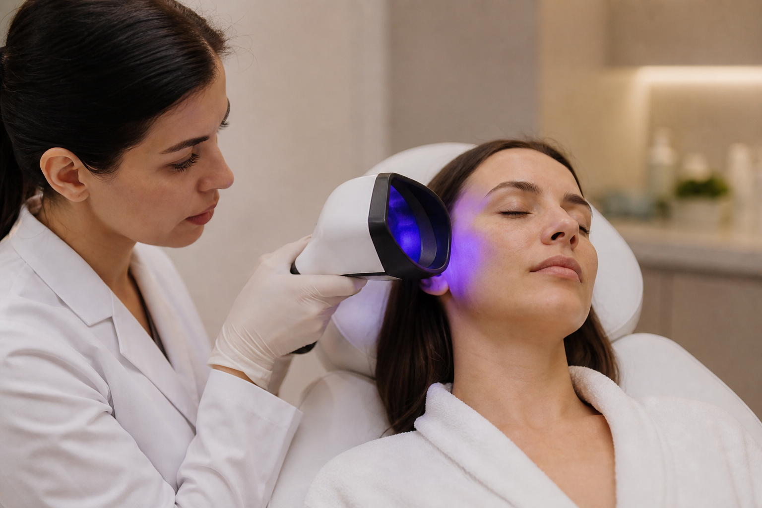
During the procedure, the room is darkened to optimize visualization of fluorescence. The lamp is positioned a few centimeters away from the skin, and the clinician systematically scans all relevant areas, noting fluorescence colors and patterns. The findings are then correlated with clinical observations to generate an informed assessment.
Following the examination, the clinician discusses the results with the patient, explaining the significance of the pigmentation patterns, possible diagnoses, and recommended next steps, which may include additional tests, dermatologic evaluation, or treatment planning.