The appearance of facial capillaries is a common concern that affects a diverse group of individuals across various ages and skin types. These visible, fine blood vessels close to the surface of the skin are sometimes described by terms such as telangiectasias or spider veins, and they frequently manifest around areas like the nose, cheeks, and chin. Understanding why facial capillaries appear is essential for those seeking to maintain an even complexion or explore appropriate cosmetic or medical interventions. This article delves deeply into the origins and causes of visible facial capillaries, clarifies the distinctions between similar vascular conditions, and provides an informative overview that supports informed decision-making when considering treatments, particularly laser procedures prevalent in advanced dermatological care.
Facial capillaries are of particular interest because their presence not only affects aesthetic appearance—often contributing to uneven redness and a blotchy complexion—but can also indicate underlying skin sensitivity or medical conditions. For individuals experiencing these vascular changes, whether sporadically due to environmental exposure or chronically owing to genetic predispositions, recognizing the causes is the first step toward effective management. Moreover, those in the realm of dermatology and cosmetic treatment benefit from a clear understanding of how facial capillaries arise to guide patient consultations and tailor vascular laser treatments that target such issues without unrealistic expectations about permanent vessel removal.
By exploring the physiological, environmental, and lifestyle factors that contribute to the visibility of facial capillaries, this comprehensive guide aims to illuminate the complexities behind these delicate vessels’ emergence. It integrates established dermatological knowledge with the latest insights into advanced laser therapies, situating the discussion within the broader context of skin health. Whether motivated by cosmetic enhancement or general skin wellness, readers will come away with a nuanced grasp of why capillaries appear on the face and what considerations matter most in managing their visibility.
What Are Facial Capillaries and How Are They Different from Other Visible Veins?
Facial capillaries, often referred to as telangiectasias in medical terminology, are tiny, dilated blood vessels near the skin’s surface that typically measure less than 1 millimeter in diameter. They manifest as thin red, blue, or purple lines and may resemble delicate spider web patterns, hence the term spider veins used colloquially. Unlike larger varicose veins or leg veins—which are commonly found on the lower extremities and tend to be thicker and more prominent—facial capillaries are exceedingly fine and superficial.
The distinction between facial capillaries and other vascular anomalies is essential for diagnosis and treatment. Telangiectasias encompass not only isolated capillaries but also specific vascular malformations involving small arteries or veins. Spider veins on the face may overlap with telangiectasias in appearance but can differ in origin and location. For example, visible veins on the legs differ in vessel size and hemodynamics and often require different laser technologies and treatment protocols compared to facial capillaries.
Understanding these differences helps clarify why certain treatments, like vascular laser procedures or intense pulsed light (IPL) therapies, vary between facial and leg vascular lesions. Furthermore, recognizing the nuances among vascular lesions fosters realistic expectations regarding the extent of improvement achievable and the likelihood of new vessel formation due to ongoing factors.
Which Factors Contribute to the Appearance of Facial Capillaries?
The etiology of visible facial capillaries is multifactorial, involving intrinsic and extrinsic elements that compromise the integrity and resilience of blood vessel walls. One significant contributor is skin type and genetic predisposition. Individuals with fair or delicate skin often have thinner dermal layers, making underlying capillaries more conspicuous and susceptible to dilation.
Environmental factors also play a critical role. Chronic sun exposure leads to photo-damage that weakens the collagen and elastin supporting facial capillaries, causing them to dilate and become visible. Similarly, exposure to extreme temperatures—both hot and cold—can induce vascular reactivity, contributing to the dilatation of these tiny vessels. For instance, regular face flushing in response to heat or cold weather or environmental irritants may exacerbate capillary visibility.
Other factors include lifestyle habits such as alcohol consumption, which dilates blood vessels transiently but can lead to longer-term vascular fragility when excessive. Smoking introduces toxins that damage blood vessels and impair circulation. Hormonal influences, including those from pregnancy, menopause, or contraceptive use, can affect vascular tone and vessel wall stability. Additionally, mechanical trauma or certain inflammatory skin conditions, such as rosacea, accelerate capillary dilation and proliferation.
Finally, aging itself contributes to capillary visibility. As skin loses thickness and elasticity over time, the protective tissue around vessels thins, rendering them more apparent. Repeated small injuries to capillaries from environmental or lifestyle triggers cumulatively lead to their dilation and permanence on the facial skin surface.
How Do Capillaries Around Specific Facial Areas Differ, and What Causes Them?
Facial vascular patterns and the propensity for visible capillaries can vary by location, influenced by anatomical and functional factors unique to each area. Around the nose, capillaries are frequently observed due to the high density of superficial blood vessels that facilitate facial thermoregulation. The nose’s prominence and continual exposure to environmental elements make these vessels particularly vulnerable to dilation from sun, temperature extremes, and irritants.
Cheek capillaries often emerge in conditions marked by chronic inflammation or vascular fragility. The cheeks host a dense network of blood vessels and are prone to damage due to frequent sun exposure and potential mechanical irritation from skincare routines or environmental factors. Additionally, people with rosacea frequently show prominent cheek vessels due to chronic facial redness and increased vascular reactivity.
Capillaries on the chin, while less common, occur typically in contexts involving hormonal fluctuations or localized skin stressors. This area, being often less exposed than the nose and cheeks, may demonstrate visible vessels in response to acne, inflammation, or hormonal changes.
Recognizing that capillary presence and severity differ by facial region aids in precise assessment and tailoring of appropriate vascular laser treatments, as different areas may respond distinctively to therapy.
What Are the Underlying Biological Mechanisms Behind Capillary Dilation?
At the microscopic level, the appearance of visible facial capillaries results from dilated and sometimes tortuous tiny blood vessels due to the weakening of vessel walls and loss of perivascular support tissue. Under normal conditions, capillaries regulate blood flow finely to provide oxygen and nutrients while maintaining skin homeostasis. However, chronic stimuli such as ultraviolet radiation, inflammation, and physical trauma provoke endothelial damage and degradation of collagen fibers that reinforce vessel walls.
The imbalance between vasodilators and vasoconstrictors at the cellular level leads to persistent dilation. Moreover, inflammatory processes trigger the release of mediators like histamine and vascular endothelial growth factor (VEGF), which promote neovascularization and vessel permeability changes. These cumulative effects thin out healthy blood vessel walls, making vessels visibly prominent through the skin’s surface.
Additionally, genetic variants affecting connective tissue proteins can predispose some individuals to fragile capillaries, exacerbating their dilation upon exposure to environmental triggers. The slow impairment of vessel integrity is often progressive but influenced by modifiable external and internal factors.
Why Is It Important to Differentiate Facial Capillaries from Other Skin Conditions?
Accurate identification of facial capillaries versus other dermatological or vascular conditions has practical importance in treatment selection and prognosis. For instance, redness attributable purely to vascular dilation differs fundamentally from erythema caused by inflammatory acne, eczema, or allergic reactions. Misdiagnosis may lead to ineffective or inappropriate therapies.
In the realm of vascular treatments, knowing whether visible lines are classic telangiectasias, spider veins, or leg veins guides the choice of laser wavelength, intensity, and procedural approach. Leg veins, often larger and deeper, require different wavelengths and sometimes different laser technologies compared to tiny facial capillaries. Misapplication can result in suboptimal results or adverse effects such as bruising or pigmentation changes.
Moreover, some rare conditions mimic facial capillaries but demand distinct medical evaluations, such as hereditary hemorrhagic telangiectasia or systemic lupus erythematosus. Thus, distinguishing these visible vessels within proper clinical context is crucial for patient safety and outcome optimization.
What Can One Expect When Considering Laser Treatment for Visible Facial Capillaries?
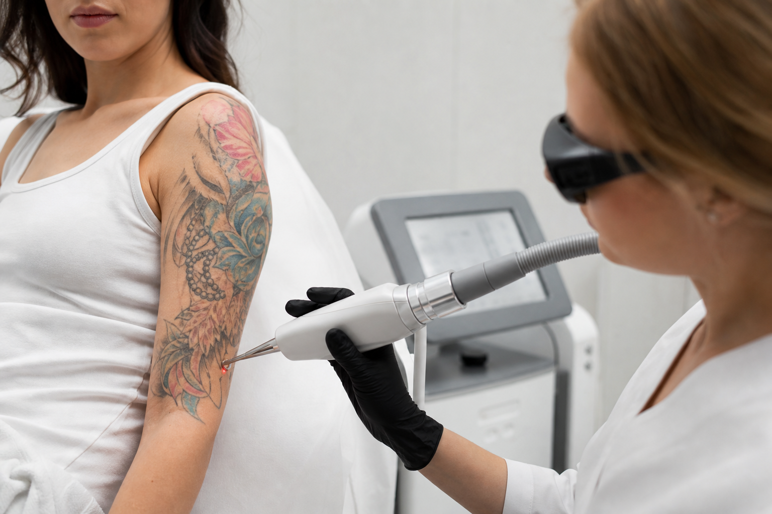
Laser treatments for facial capillaries form a cornerstone of advanced dermatological care aimed at reducing redness and vascular visibility. These procedures use targeted light to selectively heat and collapse dilated capillaries without damaging surrounding skin, leading to gradual vessel absorption by the body. Various modalities such as pulsed dye lasers, Nd:YAG lasers, and IPL have proven effective depending on vessel size, depth, and patient skin characteristics.
Patients contemplating such treatments should understand that multiple sessions are often necessary to achieve meaningful improvement. Additionally, laser therapy addresses existing visible vessels but does not guarantee that new capillaries won’t form later, particularly if causative factors persist. Hence, ongoing skin care, sun protection, and lifestyle modification are integral adjuncts to clinical treatment.
The safety profile of vascular laser treatments is favorable, but transient side effects such as mild redness, swelling, or pinpoint bruising may occur. Close consultation with a qualified dermatologist can tailor expectations, select appropriate technology, and determine the best timing considering individual vascular and skin conditions.
When Should One Seek Dermatological Advice for Facial Capillaries?
While visible facial capillaries are commonly benign and primarily of aesthetic concern, certain circumstances warrant expert consultation. Rapid onset of extensive vascular changes, symptomatic skin irritation, or the presence of associated skin symptoms such as persistent redness or flushing may signal underlying dermatological diseases like rosacea or systemic conditions that require diagnosis and management.
Consulting a dermatologist is advisable when vascular lesions interfere with quality of life, cause psychological distress, or when at-home measures fail to mitigate appearance. Specialists can perform thorough evaluations to exclude complex causes, advise on comprehensive treatment plans including laser vascular therapies, and monitor for new or worsening lesions.
Early professional engagement also helps establish a baseline skin condition and reduces the risk of progression or complications related to chronic vascular fragility.
How Do Facial Capillaries Compare to Leg Capillaries in Terms of Treatment and Appearance?
Facial and leg capillaries differ notably in both appearance and clinical management. Leg capillaries, or superficial telangiectasias of the lower extremities, often have a more reticulated pattern and larger caliber due to gravity-related venous pressure. They sometimes connect to underlying venous insufficiency, complicating treatment approaches.
Treatment modalities for leg capillaries may include sclerotherapy, which is typically not used for facial vessels, as well as specialized laser devices calibrated to penetrate deeper and target larger vessels. Facial capillaries, in contrast, are finer and closer to the surface, requiring gentler parameters and shorter pulse durations to avoid skin damage and pigmentation issues.
From an aesthetic perspective, facial capillaries are more readily visible due to thinner skin and high surface exposure, whereas leg capillaries are often concealed by clothing but can be symptomatic. Understanding these differences ensures appropriate modality selection and realistic expectations regarding outcomes and recurrence.