Solar lentigines are among the most common pigmentary changes that occur predominantly due to chronic sun exposure and photoaging. Also known colloquially as sun spots or age spots, these benign but cosmetically distressing lesions typically appear as flat, sharply demarcated brown patches on sun-exposed areas like the face, hands, shoulders, and décolletage. Their presence is often an unwelcome reminder of cumulative ultraviolet (UV) damage and underlines the importance of effective skin protection and management.
This comprehensive article serves individuals who have noticed new or increasing dark spots on their skin and are seeking to understand what solar lentigines are, why they develop, and what treatment options are available. Additionally, dermatologists, laser therapy specialists, and esthetic practitioners will find detailed guidance on diagnosis, modern treatment modalities, expected outcomes, and post-procedure care. Understanding solar lentigines is critical not only for cosmetic reasons but also because these lesions signal underlying photodamage, which can predispose to more serious dermatological conditions.
The following sections will elucidate the clinical nature of solar lentigines, their pathophysiology, when to seek expert evaluation, and the spectrum of therapeutic approaches—including the latest in laser removal techniques—along with realistic expectations, recovery considerations, and the importance of lifelong photoprotection. Where applicable, links to related topics in laser pigmentation correction within advanced laser procedures will be naturally integrated to enrich the discussion.
What Are Solar Lentigines and How Can They Be Identified?
Solar lentigines are well-circumscribed, flat, brown macules that result from localized hyperpigmentation induced by UV radiation. Unlike freckles, which tend to fade without sun exposure, solar lentigines are persistent and more prominent with age and cumulative UV damage. These lesions range in size from a few millimeters to several centimeters and often appear symmetrically on chronically exposed sites, including the dorsal hands, face, upper chest, and forearms.
Clinically, solar lentigines present as uniformly pigmented brown to dark brown patches with clear borders and a smooth surface. They do not cause symptoms such as itching or pain, which often leads to delayed presentation until their appearance becomes cosmetically bothersome. Distinguishing solar lentigines from other pigmentation disorders—like melasma, post-inflammatory hyperpigmentation, or melanocytic nevi—is critical, as management strategies differ significantly.
A confirmed diagnosis usually involves a dermatologic evaluation potentially augmented by dermatoscopy, which helps differentiate these benign lentigines from atypical or malignant pigmented lesions. Any new, changing, or irregular pigmented lesions should prompt timely dermatologic assessment to exclude melanoma or other malignancies before any cosmetic treatment is considered.
What Causes Solar Lentigines to Develop?
The primary driver of solar lentigines is chronic exposure to ultraviolet radiation—mainly UVA and UVB rays—that leads to photo-induced skin damage. UV radiation stimulates melanocytes, the pigment-producing cells in the epidermis, to produce melanin as a natural defense against DNA damage. However, excessive or prolonged UV exposure disrupts the normal distribution and degradation of melanin, causing focal accumulation and the visible pigmentation characteristic of solar lentigines.
At the cellular level, UV radiation causes oxidative stress and DNA damage in keratinocytes and melanocytes, which may result in melanocyte hyperplasia and increased melanogenic activity. Furthermore, photoaging alters the dermal matrix, weakening skin elasticity and function, which indirectly influences pigmentary changes.
Other contributing factors include genetic predisposition, advancing age, and certain hormonal influences, but these are generally secondary to UV exposure. The linkage between UV radiation and solar lentigines reinforces the critical role of sun protection in prevention.
When Should You See a Dermatologist About Pigmented Lesions?
Although solar lentigines are benign, it is crucial to undergo a thorough dermatological examination when new pigmentary spots appear, existing lesions change in color, size, shape, or texture, or when there is any irregularity or asymmetry. Suspicious lesions require dermatoscopic evaluation or biopsy to rule out malignant melanoma or other skin cancers.
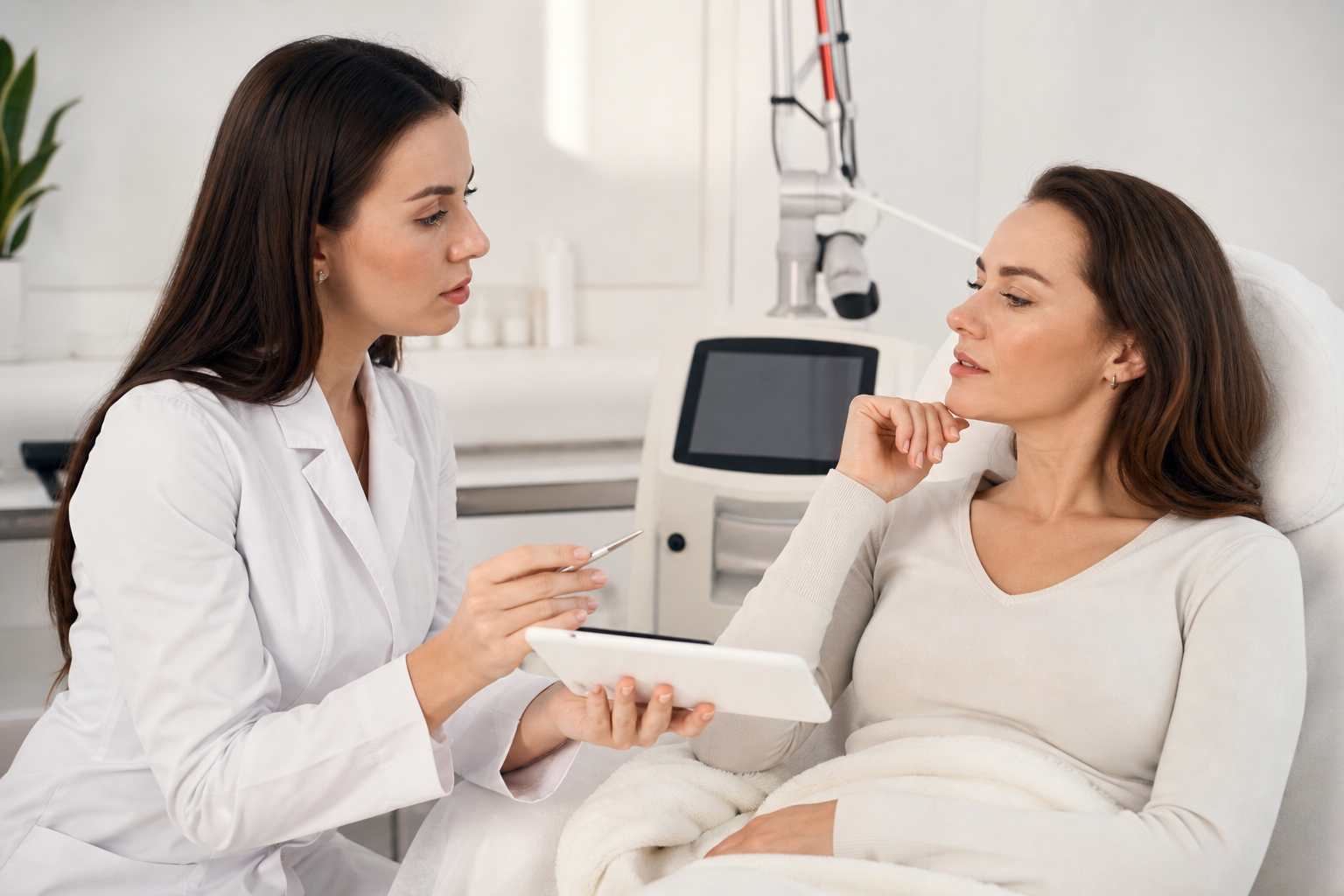
Furthermore, before undertaking any laser or cosmetic treatment for pigmentary lesions, a qualified dermatologist’s assessment is indispensable. Certain skin conditions or malignancies may contraindicate laser procedures or require alternative therapeutic interventions. Misdiagnosis or undertaking treatment without prior evaluation can lead to adverse events or delay appropriate care.
Referral to a dermatology specialist experienced in pigment disorders ensures accurate diagnosis, proper management, and safe aesthetic outcomes.
What Are the Most Effective Treatments for Solar Lentigines?
Solar lentigines can be addressed through multiple modalities tailored to lesion characteristics, skin type, patient preferences, and clinical expertise. Treatments aim to reduce pigmentation, restore a uniform skin tone, and improve overall skin quality.
Laser treatments have become the cornerstone of modern therapy due to their precision and efficacy. Q-switched lasers, Nd:YAG lasers, and fractional lasers target melanin selectively, fragmenting pigment clusters while minimizing damage to surrounding tissue. The choice of laser type and parameters depends on lesion depth, size, and patient skin phototype. Laser Removal of Sun Spots and Solar Lentigines offers an in-depth exploration of this approach within advanced laser treatments.
Other modalities include Intense Pulsed Light (IPL) therapy, which uses broad-spectrum light to target pigmented and vascular components, useful especially for mixed photoaging issues. IPL may be less precise but effective for diffuse pigmentation; a comparative discussion can be found in IPL vs Laser for Sun Spots: A Comparison.
Chemical peels employing agents like glycolic acid or trichloroacetic acid encourage exfoliation and pigment modulation, often serving as adjunctive therapies. Topical depigmenting agents such as hydroquinone, retinoids, or vitamin C help maintain results and support pigment control.
What Should Patients Expect During and After Laser Treatments?
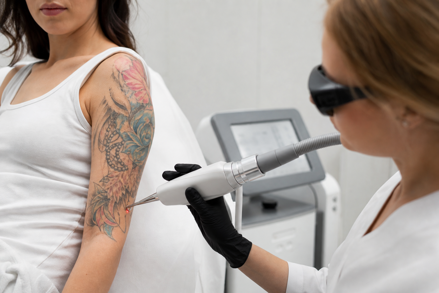
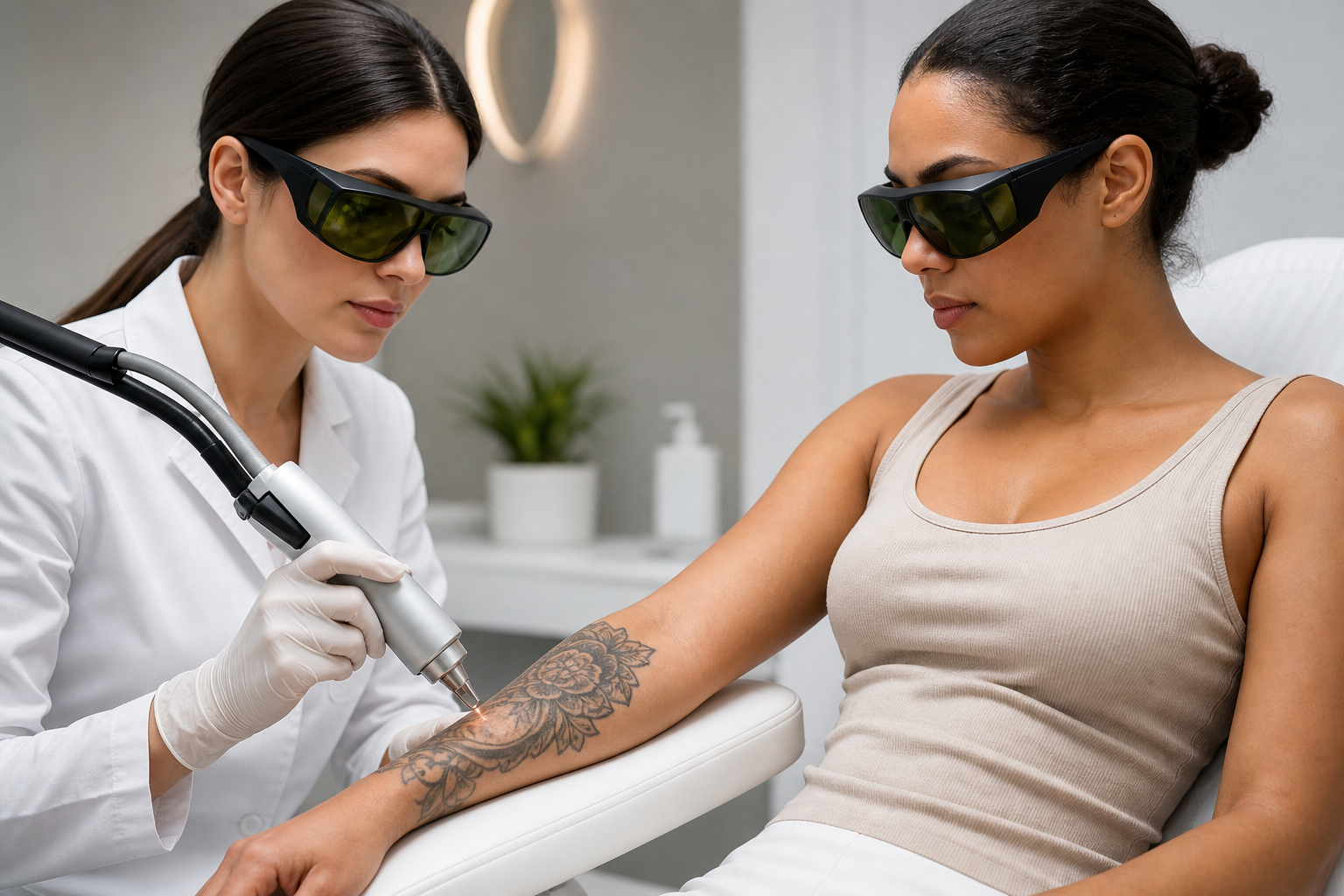
Laser sessions are generally brief and well-tolerated, though some patients may experience mild discomfort akin to snap sensations or warmth during the procedure. Anesthetic creams can be applied to enhance comfort. Multiple sessions—typically between three to five—are often necessary to achieve significant pigment clearance.
Post-procedure, patients may observe transient redness, slight swelling, or crusting in treated areas, indicating effective melanin disruption and skin healing. These expected reactions usually resolve within days to a week. Adherence to post-treatment care, including moisturization and rigorous sun protection, is vital to facilitate recovery and optimize long-term results.
Patients should be counselled that while laser therapy can provide marked improvement, it does not guarantee permanent removal as pigmentary lesions may recur with ongoing UV exposure or intrinsic skin aging processes. For details on session frequency and protocol considerations, see How Many Treatments Are Needed for Sun Spots?.
How Can One Prevent the Formation and Recurrence of Solar Lentigines?
Effective prevention hinges on meticulous photoprotection. Daily application of broad-spectrum sunscreens with SPF 30 or higher on all exposed skin remains the most crucial strategy. Protective clothing, wide-brimmed hats, and scheduling outdoor activities outside peak UV hours further reduce UV burden.
Beyond external measures, integrating antioxidant-rich skincare to combat oxidative stress and consulting dermatologists for topical agents that regulate pigmentation can enhance prevention. Educating patients on regular skin self-examination and professional skin assessments promotes early detection and intervention.
How Safe Are Laser Treatments, and When Are They Recommended?
When performed by experienced professionals using state-of-the-art equipment, laser treatments for solar lentigines are generally safe with minimal risks. Pre-treatment evaluation is critical to identify contraindications such as active infections, photosensitive conditions, or certain skin types vulnerable to adverse effects.
Laser therapy is recommended particularly when non-invasive methods have been ineffective or when pigmentary lesions substantially impact a patient’s quality of life or aesthetic satisfaction. It also forms part of a comprehensive skin rejuvenation regimen, targeting other photoaging signs like fine wrinkles, uneven texture, and vascular irregularities.
Why Is a Dermatological Evaluation Crucial Before Cosmetic Pigmentation Treatments?
Pre-treatment dermatological assessment ensures that pigmentatory lesions are properly diagnosed, weed out malignant or premalignant conditions masquerading as benign spots, and tailor therapy according to skin type and lesion characteristics. This safeguards against inappropriate treatment that could cause scarring, hypopigmentation, hyperpigmentation, or overlooked malignancies.
Comprehensive evaluation may include dermatoscopy, biopsy if needed, and consideration of patient history including sun exposure, family history, and prior treatments. The dermatologic approach integrates safety, efficacy, and patient counseling to maximize therapeutic success.